Table of Contents
Definition
Epithelial tissue is a sheet of cells joined together through the cell to cell adhesions that line the lumen of body cavities and also the outer surfaces of the body; they also line glands (serving as the secretory cells of the glands) and also line ducts of glands. When these sheets of cells form many layers, it is called multi-layered epithelium (or stratified epithelium) but when the cells form a single layer, it is called Unilayered epithelium (or Single epithelium). A single epithelial tissue is called an Epithelium and multiple epithelial tissues are called Epithelia.
What Is Epithelial Tissue?
Epithelial tissue is one of the four primary types of tissues and there are different types of epithelial tissue that are made up of diverse morphologies and functions. Epithelial tissues cover the body surfaces; they line body cavities and also form a variety of glands.
Types of Epithelial Tissues
- Simple epithelia contain one cell layer
- Stratified epithelia contain two or more layers of cells
- Transitional epithelia contain layers of cells whose morphology changes such as from squamous cells to columnar cells as you move across the layers of cells. The cellular nuclei are not at the same level.
- Pseudo-stratified epithelia the nuclei of these cells appear to be arranged in two or more layers giving the impression that the epithelium is more than one cell thick but are actually only one layer of cells with some cells being broader near the base while others are broader near the apex and their nuclei lie in the broader part of each cell and are, therefore, not in one layer
This classification of epithelia is based on the cell layers and morphology.
Simple Epithelium
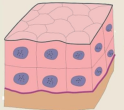
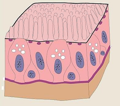
This has one layer of cells and all cellular nuclei are at the same level. There are different types of simple epithelia.
Types of simple Epithelium
- Simple squamous epithelium
- Simple cuboidal epithelium
- Simple columnar epithelium
Stratified Epithelia
This has two or more layers of cells having their nuclei at the same level. There are different types of stratified epithelia.
Types of Stratified Epithelia
- Squamous keratinized epithelium
- Squamous non-keratinized epithelium
- Stratified Cuboidal epithelium
- Stratified Columnar epithelium
- Stratified Transitional epithelium
Characteristics and Structure of Epithelial Tissue
Epithelial tissues are composed of majorly cellular components with little extracellular matrix (ECM). This structure makes it possible for cells to have cell adhesion and communication which helps them function very well. Epithelial tissues sit on top of the basement membrane this basement membrane separates epithelial tissues from underlying connective tissues.
Epithelial tissues do not have their own blood supply and only get their blood supply by diffusion from nearby loose connective tissues such as areolar connective tissue. Because of the dependency of epithelial tissues on surrounding loose connective tissues, their degree of thickness is limited. The functions and classification of epithelial tissues are based on their organization and the types of cells in epithelial tissue.
Components
- Epithelial cells these cells have a high capacity for regeneration
- Basement membrane (a combination of the basal lamina and the reticular lamina)
- intercellular junctions (cell adhesions)
Intercellular junctions (cell adhesions)
Epithelial cells are bound together very firmly and they tend to resist forces that separate them by means of intercellular junctions. There are different types of cell adhesion molecules that form intercellular junctions. These junctions are found between the cell membranes of adjacent epithelial cells and they are narrow (about 20 nm) and contain a small number of proteoglycans that are rich in cations (positive electrodes). The types of intercellular junctions and the functions of each adhesion molecule will be mentioned below.
Types of Intercellular junctions of epithelial tissues
- Zonula occludens (also known as occluding junction or tight junction or impermeable junction): these adhesion molecules help to tight the cell membranes of adjacent cells making sure that they are in contact with each other, forming a web-like seal and preventing diffusion of materials across cells thereby maintaining polarity of the cells. This type of adhesion molecule is located at the lateral and apical parts of the cell membranes.
- Zonula adherens (also called adhesion junctions): this is a band-like adhesion junction that helps to close adjacent cell membranes and resists any force that tends to separate the cells. These junctions are found below the zonula occludens.
- Desmosomes (also known as macula adherens): this helps to anchor cells together and is located below the zonula adherens.
- Gap junctions (also called communicating junctions): this keeps adjacent cell membranes in close proximity and allows the direct passage of signaling molecules between cells. They are located below the zonula adherens.
- Hemidesmosomes: these are found inside the epithelial cells and not between the cells. They are intracellular plaque similar to desmosomes but have intermediate filaments. They are responsible for anchoring epithelial tissue to the basement membrane and connective tissue thereby resisting abrasion and preventing separation between the epithelium and the connective tissue
Basement Membrane
Epithelial cells rest on a basal lamina that separates the epithelium from underlying connective tissue and provides support and a surface for the attachment of the overlying epithelium. The basal lamina is produced by the epithelial cells this basal lamina is reinforced on the connective tissue side by a layer of reticular fibers embedded in proteoglycan (known as the Reticular lamina). The basal lamina and the reticular lamina together form the Basement Membrane which is visible under a light microscope.
Types of Epithelial Cells
- Squamous epithelial cells are flat
- Cuboidal epithelial cells are cube-like
- Columnar epithelial cells are columnar in shape and have secretory functions
Epithelial Tissue Functions
- Epithelial tissue serves as a covering and lining to body surfaces thereby protecting these surfaces such as the epidermis of the skin that protects the muscles and other underlying tissues
- Epithelial tissue helps in the absorption of nutrients from the gastrointestinal tract into the bloodstream such as the intestinal lining
- Epithelial cells are responsible for secretion in glands such as the parenchymal cells of glands
- Some cells of certain epithelia have the ability to contract such as the myoepithelial cells while others have sensory ability such as specialized sensory cells of taste buds or the olfactory epithelium
In general, all substances that enter or leave the body must pass through the epithelial tissue.
Location
- Simple squamous epithelium forms the lining of blood vessels (endothelium)
- Simple squamous epithelium forms the serous lining of the lungs (pleura), of the heart (pericardium), etc.
- Simple cuboidal epithelium forms the covering of the ovary and the thyroid
- Simple columnar epithelium forms the lining of the intestines and gall bladder
- Transitional epithelium forms the lining of the trachea, bronchi, and nasal cavity
- The pseudostratified columnar epithelium is found in some parts of the auditory tube, the ductus deferens, and the male urethra (membranous and penile parts)
- Keratinized stratified squamous epithelium forms the epidermis of the skin
- Non-keratinized stratified squamous epithelium forms the lining of the mouth, esophagus, and larynx.
- Stratified cuboidal epithelium is found in sweat glands and developing ovarian follicles
- Stratified transitional epithelium is found in the bladder, ureters, and renal calyces
- The stratified columnar epithelium is found in the conjunctiva of the eyes


