Table of Contents
What is Areolar Connective Tissue?
Areolar connective tissue is a loosely arranged connective tissue that is widely distributed in the body and contains collagen fibers, reticular fibers, and a few elastic fibers embedded in a thin, almost fluid-like ground substance. The areolar connective tissue is a subtype of loose connective tissue.
It is the commonest type of connective tissue in the human body and it is found in most organs of the body and in other tissues. Sometimes, the Areolar connective tissue is also known as loose connective tissue because when viewed under low magnification with a microscope, it is found to be made up mainly of bundles of loosely arranged fibers that appear to enclose large spaces called Areolae and it is the commonest type of loose connective; however, there are other types of loose connective tissues such as the reticular tissue, the mesenchyme, Mucoid connective tissue, and the adipose connective tissue.

Areolar Connective Tissue Functions
- This tissue functions mainly in binding organs and their components together providing elasticity when stretched
- It forms helices around long axes of expandable tubular structures such as blood vessels, ducts of glands, and gastrointestinal tract thereby providing support and cushion to these structures
- This tissue supplies blood to the nearby epithelial tissue
- It contains leucocytes (white blood cells) that help in fighting infections and the space of the areolar tissue helps these cells to move about freely and easily find infectious cells.
Structure and Histology
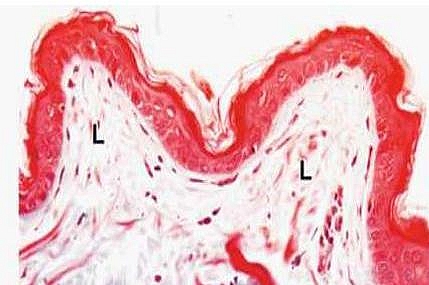
In areolar connective tissue, the fiber bundles are loosely arranged with wide spaces in between them. It contains the random distribution of much ground substance, little collagen, and a variety of cells together with blood vessels that are numerous. The areolar connective tissue appears different in various organs and may even appear compact when the areolar tissue of the skin is viewed with a microscope making it difficult to be distinguished from dense irregular connective tissue.

Areolar connective tissue is best identified under the microscope due to its lack of structure the fibers are randomly arranged. If you find a random arrangement of tissue under the microscope with spaces, it is most likely the areolar tissue you are viewing.
Components and Composition of the Areolar Connective
- It contains cells: fibroblasts, white blood cells, and mast cells
- It contains fibers: elastin, collagen, and reticular fibers
- Areolar tissue also contains the ground substance which is like a fluid matrix in which are other components of this tissue – cells and fibers
The Cells
- Macrophages these cells phagocytize (eat up) pathogens or germs or infectious cells found in the body
- Mast cells these cells release histamine during hypersensitivity reactions
- Fat cells (adipocytes) secrete lipids
- Plasma cells– they are involved in immune mediation by producing antibodies
- Fibroblasts secrete the fibers in areolar tissue. These are the most abundant cell types found in areolar tissue
The Fibers
- Collagen fibers
- Elastin fibers
- Reticular fibers
Areolar Tissue Location in the body
- Commonly found under epithelial tissues
- Found in the dermis of the skin
- They are also found in the lamina propria of the gastrointestinal tract and also of the respiratory tract.
- It is found in the layers surrounding glands and their ducts
- In the mucous membrane of the urinary tract and reproductive tract.