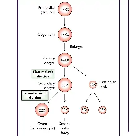
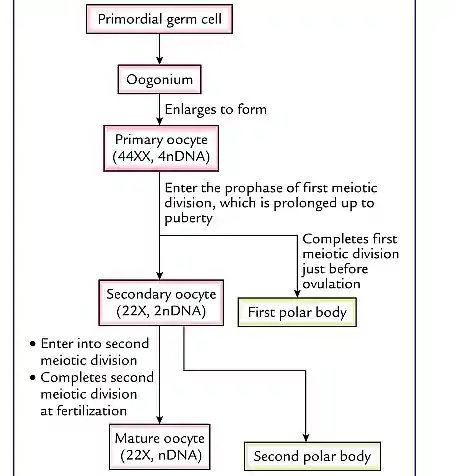
Oogenesis is also called Ovogenesis, and it is the process whereby oogonia (immature oocytes) are transformed into mature oocytes. The process of Oogenesis begins before birth and is completed after puberty. Oogenesis even after puberty still continues to menopause (permanent cessation of menstrual bleeding associated with the menstrual cycles). Oogenesis and Spermatogenesis are forms of Gametogenesis (formation of mature male and female gametes from primordial germ cells). Oogenesis occurs in the ovaries of females.
The Primordial germ cells divide by mitosis to form a large number of Oogonia with each oogonium enlarging to form a primary oocyte. Thus Oogenesis ultimately produces one primary oocyte forming only one ovum with 22 autosomes and one X chromosome; three polar bodies each with 22 autosomes and one X chromosome are formed. Oogenesis is accompanied by the development and growth of the follicles.
Table of Contents
Oogenesis comparison with Spermatogenesis
Unlike in males during spermatogenesis, there is no formation of primary oocytes after birth in females. The primary oocytes remain inactive in the ovarian follicles until puberty. As a follicle matures, the primary oocyte enlarges and, shortly before ovulation, completes its first meiotic division to give rise to a secondary oocyte and the first polar body but this stage is different from the corresponding stage of spermatogenesis, in that the division of cytoplasm is unequal in this stage in oogenesis because the secondary oocyte receives almost all the cytoplasm with the first polar body receiving very little. The Polar body is a small, non-functional cell that soon degenerates.
At ovulation, the second meiotic division is started by the nucleus of the secondary oocyte and is halted at metaphase and only completed when if a sperm cell fertilizes the secondary oocyte with most of the cytoplasm being retained by the fertilized oocyte and the other cell, the second polar body (a small nonfunctional cell) soon degenerates; maturation of the oocyte is completed when the polar body is extruded.
Number of Oocytes in the female ovary
There are about two million primary oocytes in the ovaries of a newborn female, but most regress during childhood so that by adolescence no more than 40,000 oocytes remain of these, only about 400 become secondary oocytes and are released at ovulation during the reproductive period with a few of these oocytes becoming fertilized and mature.
Stages of Oogenesis
The stages of Oogenesis can be grouped into two phases: Prenatal and Postnatal stages of maturation of oocytes.
Prenatal Maturation of Oocytes
During early fetal life, Oogonia proliferate by mitosis and then enlarge to form Primary Oocytes before birth. As primary oocyte forms, connective tissue cells surround it and form a single layer of flattened, follicular epithelial cells and together the primary oocyte and the enclosing layer of cells form a Primordial follicle.
As the primary oocyte enlarges during puberty, the follicular epithelial cells transform into cuboidal cells and the columnar cells thereby forming a Primary follicle . The Primary oocyte is usually surrounded by acellular glycoprotein covering known as the Zona pellucida which appears as a regular mesh-like appearance with intricate fenestrations on electron microscopy.
Primary oocytes always begin the first meiotic division before birth, but completion of the phase of prophase does not occur until adolescence as a result of the substance called oocyte maturation inhibitor that is secreted by the follicular cells surrounding the primary oocyte. This substance keeps the meiotic process of the oocyte arrested until adolescence.
Postnatal Maturation of Oocytes
Shortly before birth, all primary oocytes have started the prophase stage of meiosis I, but instead of proceeding into metaphase, they enter the diplotene stage, a resting stage during prophase that is characterized by a lacy network of chromatin. The Primary oocytes remain dormant in prophase and do not finish their first meiotic division until puberty due to the effect of oocyte maturation inhibitor(OMI), a substance secreted by follicular cells.
The total number of primary oocytes at birth is estimated to vary from 700,000 to 2 million with most undergoing atrophy during childhood leaving approximately 400,000 at the beginning of puberty with fewer than 500 being ovulated. Some oocytes that reach maturity late in life have been dormant in the diplotene stage of the first meiotic division for about 40 to 45 years or more before ovulation this long duration of the first meiotic division which can last for an average of 45 years may be responsible for the relatively high frequency of meiotic errors leading to congenital anomalies observed with increasing maternal age. These primary oocytes that are halted in the stage of prophase (diplotene stage) are vulnerable to environmental agents such as radiation.
At the start of puberty, one follicle usually matures and the oocyte is released via a process known as Ovulation. This process is usually inhibited medically by the use of oral contraceptives.
Important Facts about Oogenesis
- The primary oocyte enters into the first meiotic division before birth and completes it at puberty just before ovulation.
- Primary oocytes are not formed again after birth, this is the reason why every female has a specific number of oocytes (ova, eggs) that are released monthly through a process called ovulation.
- The secondary oocyte enters the metaphase stage of the second meiotic division at the time of ovulation and continues till fertilization. If fertilization occurs, then the secondary oocyte completes its meiotic division
- At birth, the ovary contains about two million germ cells with most of them degenerating and only about 40,000 oocytes are left in the ovary at puberty.
- Oogenesis involves both Mitosis and Meiosis as the proliferation of Oogonia occurs by Mitosis and the other stages of oogenesis are then completed by meiosis.


