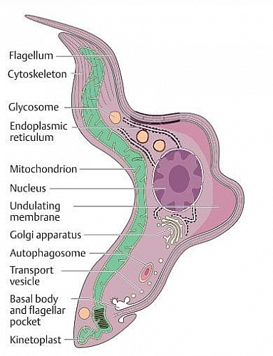
Trypanosoma Cruzi is a member of a phylum, called Sarcomastigophora in taxonomy. It is a protozoan that causes a disease called Chagas Disease or American Trypanosomiasis (this disease is different in pathogenesis and epidemiology from African Trypanosomiasis).
Table of Contents
- Trypanosoma Cruzi Epidemiology
- Trypanosoma Cruzi Transmission
- Species of Reduviid bugs (vectors of Trypanosoma cruzi)
- Trypanosoma cruzi Pathogenesis
- Trypanosoma Cruzi Morphology
- Trypanosoma Cruzi Life Cycle
- Trypanosoma Cruzi Lab. Diagnosis
- Trypanosoma Cruzi Treatment
- Prevention of Trypanosoma cruzi infection
Trypanosoma Cruzi Epidemiology
Trypanosoma Cruzi is found majorly in South America, Central America and some few regions in North America (Texas and Mexico) these are the endemic areas. Trypanosoma Cruzi infection is the major cause of heart failure in South America.
Trypanosoma Cruzi Transmission
- Reservoirs of Trypanosoma cruzi include wild animals such as opossums, rodents and armadillos. The vector for the transmission of Trypanosoma cruzi infection is the Reduviid bug (it is also called the kissing bug or assassin bug). This bug transmits Trypanosoma cruzi infection when it feeds on humans at night while they sleep; it will then defecate as it feeds; the Trypanosomes are found in the feces and are introduced into the body when the fecal matter is rubbed accidentally into the bite wound.
- Oral transmission can also occur when bite occurs near the lips or the fecal matter gets into the mouth. Blood transfusion with infected blood also transmits the infection too.
- Diaplacental infection may occur when a pregnant women infects the fetus
- Transmission of trypanosoma cruzi infection may also organ transplants (this is rare)
Species of Reduviid bugs (vectors of Trypanosoma cruzi)
- Triatoma infestans
- Triatoma brasiliensis
- Triatoma dimidiate
- Triatoma sordida
- Rhodnius prolixus
- Panstrongylus megistus
Together, these bugs are called Triatomines.
Trypanosoma cruzi Pathogenesis
When triatoma (reduviid bug) takes a blood meal, it excretes the feces from which the trypomastigotes infect humans through skin lesions such as abrasions, bug bite or even through the mucosa of the mouth or the conjunctiva of the eyes. Once in the human body, the parasites are taken up by macrophages or they just invade other cells such as cardiac muscles, skeletal muscles or smooth muscles; the parasites also invade neuroglial cells. In the cells of the host, the parasite transforms into amastigote and then multiplies by binary fission in the cells of the host. The Cells filled with up to 500 trypanosoma cruzi parasites are called pseudocysts. After about 5 to 6 days, the parasites develop into the epimastigote form and then back to the trypomastigote form and they return to the bloodstream, whereupon the cell infection cycle is repeated.
Trypanosoma Cruzi Morphology
The Trypanosoma cruzi parasite in the blood of infected hosts measures about 16 to 22 micrometer long and has a pointed posterior end and a large kinetoplast. Multiplication does not take place in the blood but in the host cells and these intracellular forms are called amastigotes (they measure about 1.5 to 4.0 micrometer long and are highly multiplicative)
Trypanosoma Cruzi Life Cycle
- The life cycle of trypanosoma cruzi begins when the reduviid bug (or Triatoma or cone-nose bug) ingests trypomastigotes in the blood of the reservoir host (Humans).
- In the intestine of the reduviid bug, trypomastigotes first change into intensively multiplying epimastigotes in order to multiply and then differentiate back into trypomastigotes. This takes a period of 6 to 7 days before being excreted in the feces.
- When the bug bites again, the site is contaminated with feces containing trypomastigotes, which are introduced into the blood of another person and they then transform into non-flagellated amastigotes within cells of the host (in human cells).
- Many cells are affected such as myocardial cells, glial cells and reticuloendothelial cells
- To complete the Trypanosoma cruzi life cycle, the amastigotes in the host cells differentiate again into trypomastigotes, which then enter the blood and are taken up by the reduviid bug
Trypanosoma Cruzi Lab. Diagnosis
- Diagnosis can be made by making a blood smear and using microscope to view the trypanosomes this can be seen in acute infection and is not effective in chronic infection
- Chronic trypanosoma cruzi infection, the parasites may be detected by xenodiagnoses whereby an uninfected reduviid bug is allowed to feed on the patient, and the insect gut is then examined for parasites after some weeks; if trypanosomes are found, then the person has chronic infection.
- Serological tests can also diagnose both the acute and chronic trypanosoma cruzi infection
Trypanosoma Cruzi Treatment
- Nifurtimox is the drug of choice for treating Trypanosoma cruzi acute infection
- Benznidazole can also be used with a cure rate of about 80% in acute infection
Prevention of Trypanosoma cruzi infection
- Use of insecticides in the rooms to kill reduviid bugs which serve as vectors
- Use of bed nets impregnated with pyrethroid
- Use of insect repellent creams
- Improve housing conditions such as avoid making thatched houses and mud houses
