Table of Contents
- What is Leukaemia?
- Functions of the Blood cells affected by Leukaemia
- Types of Acute Leukaemia
- Differences between Acute Lymphoblastic Leukaemia (ALL) and Acute Myeloid (myelogenous) Leukaemia (AML)
- Risk Factors and Causes of Acute Leukaemia
- Clinical Symptoms and Signs of Acute Leukaemia
- Diagnosis of Leukaemia
- Treatment of Leukaemia
What is Leukaemia?
Leukaemia is simply the cancer of the blood. Just like any other cell of the body, blood cells can become malignant and cannot be controlled by the normal body regulatory mechanism. This means that these Leukemic cells continue to proliferate in the blood without control. The harmful effect of this is that the cells do not perform the normal functions of blood cells; as a result of this, Leukaemia makes the normal cells of the blood become very few and the symptoms and signs of Leukaemia are due to insufficient normal cells of the blood.
Acute Leukaemia affects all types of blood cells which are red blood cells, white blood cells and Platelets. The white blood cells have different forms such as Neutrophils, Lymphocytes, Macrophages, Basophils, and Eosinophils. Each of these blood cells has different functions in the body. To understand Leukaemia, you have to know the functions of the various blood cells which are outlined briefly below.
Functions of the Blood cells affected by Leukaemia
- Red blood cells: carry oxygen to the body tissues. A deficiency of this causes a condition known as Anemia.
- Lymphocytes help to fight infections. Deficiency of this leads to infections
- Eosinophils also help to fight infections especially of parasites and inflammation
- Basophils are involved in allergic reactions as a response to foreign substances that are not compatible with the body and also in inflammatory reactions.
- Platelets help in blood clotting and deficiency of these cause easy bleeding
- Macrophages clear microorganisms in the body
As outlined above, any reduction in these normal cells by Leukaemia leads to the symptoms and signs we see in Leukaemia. Leukemic cells may look like any of the normal cells mentioned above but do not function to help the body but rather cause problems to the body.
Leukaemia normally affects the cells that generate other blood cells. These generating cells are medically known as Hematopoietic Precursor cells.Acute leukemia is, at the very least, a hematologic urgency, and it is sometimes a true emergency (if the patient has leukostasis, sepsis, Disseminated Intravascular Coagulopathy – DIC).
Types of Acute Leukaemia
- Acute Myeloid (myelogenous) Leukaemia (AML)
- Acute Lymphoblastic Leukaemia(ALL)
Differences between Acute Lymphoblastic Leukaemia (ALL) and Acute Myeloid (myelogenous) Leukaemia (AML)
- ALL is more common in children while AML is much more common in adults
- First peak age for ALL is at 3-4 years while AML increases with age
- 2nd peak age of ALL occurs in the elderly
- Out of 100 children with ALL, about 70 can be cured but only a few having AML can be cured
Risk Factors and Causes of Acute Leukaemia
- Ionizing radiation: following irradiation of any part of the body, one stand a chance of having ALL, AML or CML but it does not increase the chance of having CLL. Sometimes, it takes about 5 years after the radiation for one to have Leukaemia.
- Benzene containing drugs
- Drugs used in Chemotherapy
- Other drugs such as Cyclophosphamide, Melphalan, epipodophyllotoxins (eg. etoposide)
- Human T cell Leukaemia (HTLV-1)
- Human T-cell Lymphoma Virus
- Downs syndrome
- Fanconis anemia
- Blooms syndrome
- You are at increased risk if any of your sibling has Acute Leukaemia
- Myelodysplastic syndromes
- Aplastic anemia
- Multiple myeloma
- Waldenstrms macroglobulinemia
- Paroxysmal Nocturnal Hemoglobinuria (PNH)
Most patients with acute leukaemia do not have an identifiable cause
Clinical Symptoms and Signs of Acute Leukaemia
- Anemia: due to reduced red blood cells in the blood and the person will have fatigue and become pale
- Low Platelets in the blood (Thrombocytopenia): this will manifest with easy or spontaneous bleeding and spontaneous bruising
- Reduced Neutrophils in the blood (Neutropenia): this will cause infections and sepsis sets in.
- There could be infiltration the Leukaemic cells into other organs of the body such as Liver, Spleen, Lymph nodes and others;
- There would be swelling of lymph nodes around the neck, arm pit and other parts of the body. Lymph nodes swellings are felt like peanuts under the skin. This condition is known medically as lymphadenopathy.
- Enlargement of the liver and spleen may occur (Hepatosplenomegaly)
- Swelling of the gums of the mouth with easy bleeding even while brushing your teeth
- Bone pains or Tenderness
- Leukaemia cutis
- Chloromas
- Leukostasis giving rise to stroke, pulmonary infiltration.
- Fever
- Sweating is common
- There may be weight loss but it is uncommon
Diagnosis of Leukaemia
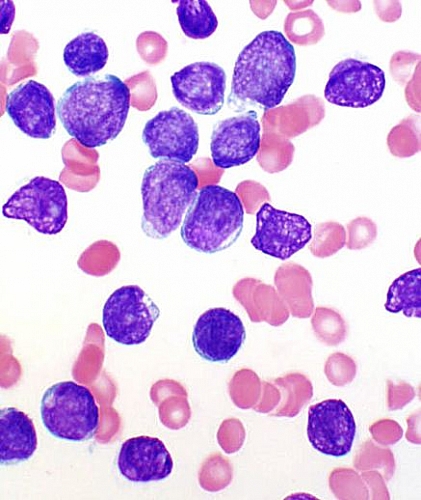
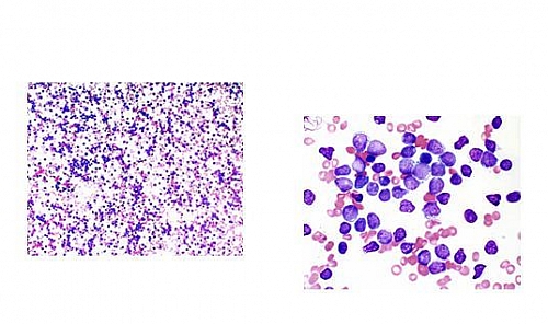
The diagnosis of Leukaemia is carried out in the laboratory and a staining agent is used to stain the blood collected from the patient. It is then viewed under the microscope. The appearance of the cells under the microscope helps to distinguish the type of Leukaemia that the person has.
Acute Lymphoblastic Leukaemia (ALL) appearance in the blood using Microscope
- Proliferation of lymphoblasts
- Small cells with indistinct nucleoli
- High nuclear/cytoplasmic ratio
- Fine chromatin
Features of Acute Lymphoblastic Leukaemia in the bone marrow using a Microscope
- Hypercellular marrow
- Excess number of blasts (Acute Leukaemia is diagnosed when over 20% of nucleated cells in the bone marrow are blasts.)
Acute Myelogenous Leukemia
AML occurs as a result of proliferation of immature myeloid cells. It primarily occurs in adults and it develops in some patients undergoing chemotherapy, radiotherapy, or other risk factors mentioned above.
Treatment of Leukaemia
Leukaemia treatment basically involves Chemotherapy or Bone Marrow Transplant. Treatment may involve a Pediatrician, Hematologist, Oncologist, Social workers, Psychologist and Srgeons.
Chemotherapy involves 3 stages which are Remission induction, consolidation and maintenance.
Remission Induction
The aim of this therapy is to eradicate the Leukaemic cells from the bone marrow. Drugs used include: Asparaginase, Prednisolone and Vincristine (Onconvin). This treatment usually last for about 4 to 6 weeks. Intrathecal therapy can be given using Methotrexate, Hydrocortisone, and Cytosine Arabinoside
Consolidation phase of Leukaemia treatment
This takes about 4 to 8 weeks and Vincristine and Prednisolone are used.
Maintenance Phase
This treatment can last for 2 to 3 years and involves using 6-Mercaptopurine, Methotrexate, Vincristine and Prednisolone.
Definitive treatment of Leukaemia
The lasting cure is bone marrow transplant and this could be Autologous transplantation (the cells of the patient are harvested during the remission phase and later used for transplant) or Donor transplantation (where a donors cells are harvested and transplanted to the recipient).
Supportive treatment in Leukaemia
Any other associated complication or disease is also treated. If there is infection or Tumor lysis syndrome, treatment is carried out. If the patient is in need of blood transfusion, that is also done.


