Table of Contents
Computed Tomography Scanning (CT SCAN)
Computed tomography is not as readily available as ultrasound, it is costly and is often a second line investigation. It is however the imaging of choice in abdominal trauma and for staging tumours. It is used for the following disease conditions.
Uses of CT SCAN in diagnosis of Abdominal Disease Conditions
- Pancreatitis. The pancreas lies posteriorly and is often obscured by bowel gas on ultrasound examination in the acute stage. Also, a post contrast CT study shows well the amount of remaining viable pancreatic tissue.
- Trauma. Ultrasound is often used as a first line but if this is negative and injury is strongly suspected CT should be performed. It is difficult with ultrasound to demonstrate bowel injury or pancreatic trauma unless a pseudo cyst is present.
- Tumour staging. Computed tomography shows nodes very clearly and demonstrates the extent of other organ involvement.
- Certain liver masses. It helps to characterise liver masses when ultrasound is unsure e.g. haemangiomas. Sometimes metastases show on CT and not on ultrasound and vice versa.
- Aetiology of abdominal mass, sometimes it is not possible to say which organ a mass has arisen from on ultrasound and in these cases CT is often helpful.
- Suspected abscess collection when ultrasound is negative.
- Elderly patient: Occasionally for a suspected large bowel lesion in an elderly patient who cannot co-operate with a barium enema.
Nuclear Medicine
This involves the use of radioisotopes in the diagnosis and treatment of diseases. It can detect micro-metastasis of cancer and is highly sensitive.
Uses of Nuclear Imaging in Abdominal diseases
Nuclear medicine, when available, is useful for the following:
- Meckels diverticulum. This causes bleeding from the bowel and if it is lined with gastric mucosa it takes up the isotope, showing as a small hot spot.
- Biliary tract. Sometimes helpful in assessing function of the gallbladder. It is called a HIDA scan
- Bleeding from the bowel when other tests have been unhelpful. Red blood cells are labelled with a radioactive isotope. If bleeding is occurring it may show as areas of radioactivity in the bowel.
Magnetic Resonance Imaging (MRI)
This involves the use of magnetic forces to help in disease diagnosis. MRI is safer than CT Scan and X-ray of abdomen as the use ionizing radiations that may predispose to tumors. It is however costly. The role of MRI is still being evaluated. It is little used for general abdominal conditions but good for specific conditions such as liver haemangiomas, staging tumours involving the uterus, bladder, and prostate. It is also good for imaging the bile ducts and pancreatic duct. It is unlikely to be available in a third world setting and even if it is, the old machines are used that may not take clear images of the abdominal conditions and even worst when the patient may not obey commands such as unconscious patients or babies.
ULTRASOUND Scanning
This makes use of sound waves in diagnosis of disease conditions. It is safe to use as it does not use ionizing radiations. It is cheap and can detect so many disease conditions of the abdomen.
Uses of Ultrasound scanning in abdominal diseases
Ultrasound is a first line investigation for:
- The biliary tract
- The liver and spleen
- Renal Tract
- Masses
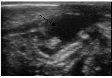
- Abscess
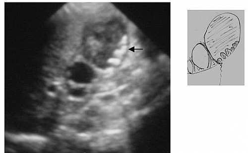
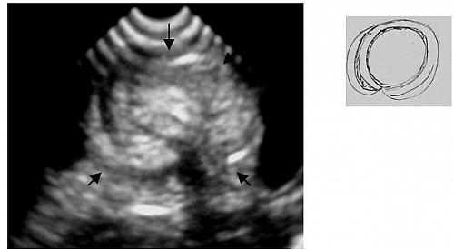
- Intussusception
- Ectopic pregnancy/ ovarian pathology/fibroids
- Ascites
- Infantile pyloric stenosis, if local expertise available
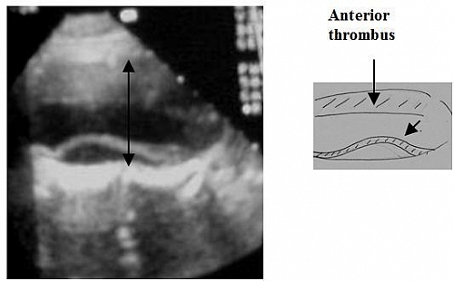
- Aortic aneurysm
- Pancreas (if no CT)
- Trauma if no CT or patient very ill
Ultrasound is also useful in:
- Inflammatory bowel disease shows as bowel wall thickening
- Appendicitis, especially when the use of MANTRELS Score is equivocal or complication such as appendiceal abscess is suspected


