Table of Contents
What is Barium Enema Test?
A barium Enema study is used to detect and locate abnormalities along the colon or Large intestines. It is a procedure that makes use of a contrast medium, Barium, in order to make the large intestine visible on x-ray film.
Use of Barium Enema to study conditions of the Large bowel
Symptoms such as altered bowel habit, rectal bleeding, abdominal pain, weight loss, and anaemia may indicate colonic disease. Colonoscopy and barium studies are complementary and equally useful but the method of investigation used depends on the local circumstances. In many countries, a double contrast barium enema is combined with a flexible sigmoidoscopy in all patients with symptoms suggestive of large bowel pathology.
Barium studies require full bowel preparation and a double contrast technique is routine, unless the patient has acute inflammatory bowel disease or obstruction. In the latter two cases, the use of water soluble contrast is preferred.A fairly high-density barium is run into the bowel per rectum as far as the hepatic flexure. Buscopan is given intravenously and most of the barium drained back. Air is then introduced by means of a Higginsons syringe causing the remaining barium to reach the caecum, the colon to fully distend with air while the mucosa is coated with barium.
Abnormalities (disease conditions) seen on barium enema examination
Redundant loop of sigmoid
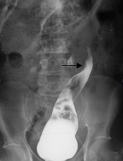
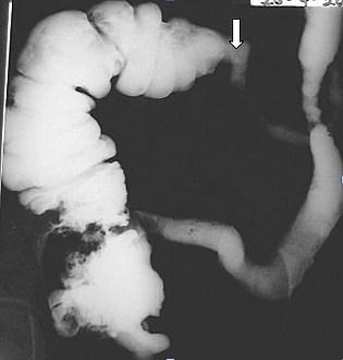
This is a normal variant and is common in Ghana. It predisposes to volvulus, when the loop rotates about its axis becoming obstructed. Unrelieved it may lead to bowel infarction and perforation. On barium enema, the colon is obstructed at the level of the volvulus and the contrast column tapers to give a birds beak or twisted ribbon appearance.
Barium Enema for Colon Cancer
Carcinoma can occur anywhere in the colon but is commonest in the rectosigmoid area. It may develop from a polyp and present as a filling defect or it may infiltrate the bowel wall appearing as a stricture. The first presentation may be large bowel obstruction. Occasionally it may penetrate into adjacent structures such as the bladder and present as a vesico-colic fistula.
Polyp
Polyps are localised mass lesions arising from the colonic mucosa. They protrude into the lumen and may have a flat broad base (sessile) or be pedunculated on the end of a stalk. They occur anywhere in the colon. The majority are benign, especially the small or pedunculated ones. Sessile polyps are pre-malignant and the object of a double contrast study is to detect polyps before malignant transformation has occurred. Multiple polyps occur in the hereditary conditions of familial polyposis coli and Peutz-jeghers syndrome. The polyps of the former have malignant potential whereas the polyps in the latter are always benign. Pseudo-polyps may occur in long- standing inflammatory bowel disease due to areas of mucosal hypertrophy.
Diverticular disease
Relatively uncommon in Africa, it is very common in the Western world and often leads to complications. The smooth muscle hypertrophies with pouch like protrusions between the thickened fibres. The mucosa and submucosa herniate through sites of weakness in the bowel wall. The sigmoid is the most frequently involved area but diverticulae may arise anywhere and are not uncommon in the caecum. Complications include acute inflammation with pericolic abscess, colonic perforation, fistula formation especially into the bladder and haemorrhage.
Inflammatory bowel disease
This is characterised by diffuse mucosal changes due to oedema and ulceration. It may affect the whole colon or only part of the colon. If due to Crohns disease, the distal ileum and caecum are commonly involved. A barium enema in the acute setting is seldom indicated and may be contraindicated because of the danger of perforation. A plain abdominal film may show a dilated colon outlined by air, toxic dilatation, which is an absolute contraindication to barium enema. Ulcerative colitis and Crohns disease are common in the Western World but in Africa an infective aetiology, such as Salmonella, Shigella, or Amoebiasis is more common. In the acute phase, the large bowel will show mucosal irregularity and small ulcers projecting from the wall. In the chronic stage there may just be generalised narrowing of the lumen and loss of haustration giving a pipestem appearance.
Intussusception
This occurs when part of the bowel invaginates on itself. The proximal bowel becomes invaginated into the lumen of the distal bowel. This may occur because of a localised lesion such as a polyp or carcinoma, which is carried by peristalsis along the bowel. As it is attached to the wall, it carries the proximal bowel with it. Most commonly however it occurs in infants between the ages of 3 months and 2 years when an inflamed Peyers (lymphoid) patch in the distal ileum is often the cause. As the bowel is carried down within the distal lumen the blood supply becomes impeded, oedema occurs and the bowel may become necrotic. It is nowadays usually diagnosed by ultrasound but if local expertise is not available, a barium enema can be performed. Sometimes it is possible to reduce an early intussusception in children by barium enema if fluoroscopy and local expertise are available. The appearances on barium enema show either as an abrupt filling defect with complete obstruction to flow, or a coil spring appearance with a little barium outlining oedematous folds.
Hirschsprungs
This disease presents in children. The patient gives a history of severe constipation from an early age due to an aganglionic segment of large bowel, which will not distend. There may be considerable abdominal distension. The aganglionic segment may occur anywhere but is usually in the distal large bowel, in the sigmoid region. Sometimes it is very low, in the rectum. If this is the case, it may not be demonstrated on barium study as the enema tube will be inserted beyond it. The appearance on imaging is that of a distal small calibre lumen with narrowing which suddenly changes to a very dilated proximal colon loaded with faeces. This is now more commonly diagnosed by rectal biopsy, as the aganglionic segment may be too low to demonstrate on barium enema. It is important not to fill the proximal dilated bowel with barium as impaction may occur. Water soluble contrast may be used instead of barium. Only a limited examination is necessary, just to show the transition in calibre and confirm the diagnosis.
Pseudo obstruction of the large bowel
Distension of the large bowel may occur in the absence of obstruction. Plain films show progressive dilatation of the colon, resembling a mechanical obstruction. It may occur in-patients who are severely ill from diseases such as pneumonia or in elderly patients who are bed ridden. It may also occur in patients with myxoedema or patients on antidepressant drugs. A barium or water-soluble enema may be necessary to exclude an obstruction.
Complications/Side Effects of Barium Enema Procedure
Although barium is a much safer contrast agent than the iodine dye based contrast media given intravenously, complications occasionally occur. The risk of developing complications can be reduced by good clinical practice and appropriate preparation of patient. Care should be taken while carrying out this technique to reduce the risk of developing these complications.
- Rectal perforation may occur with a low rectal tumour or severe proctitis when the tube is inserted for barium enema.
- Impaction of barium may occur when there is a stricture causing a degree of large bowel obstruction e.g. in Hirschsprungs
- Obstruction of the large bowel may be precipitated by a barium meal examination if there is already a sub-acute obstruction present. Water is absorbed from the barium solution in the bowel, which becomes thicker and harder. Even in-patients with no bowel abnormality it usually causes a degree of constipation.
- Myocardial infarction. This is a rare complication but distension of the large bowel may cause cardiac irregularities or angina. Occasionally it precipitates an acute infarct and barium enema is contraindicated in severe angina.
- Vaginal instillation. If the rectal tube is inserted into the vagina instead of the rectum barium will outline the uterus and fallopian tubes. If these are patent, peritonitis may occur due to spillage of barium into the peritoneal cavity.
Contraindications to performing a barium enema
- Acute toxic dilatation a transverse colon diameter of 5cm is critical for impending perforation in inflammatory bowel disease.
- Post rigid sigmoidoscopy with full thickness biopsy. Allow 2-3 days interval.
- Low rectal tumour it will often prevent the insertion of the rectal tube. Rectal tumours are best diagnosed on sigmoidoscopy.
- Severe Angina


