Table of Contents
What is Barium Meal?
Barium meal is a procedure that helps in the diagnosis of different disease conditions. This technique makes use of a medium known as “Barium”, which serves as the contrast that helps to outline the intestinal tract up to the level of the small intestine (follow through) as against a barium swallow that outlines the esophagus and the stomach only. X-rays are used and the barium appears whitish in radiographic images. Below are indications for use of barium meal and barium follow through.
Abnormalities seen on barium meal examinations (Indications for Barium Meal)
Gastric ulcer
Most commonly seen on the lesser curve but may arise anywhere. Barium collects in the ulcer crater and when seen en face on a double contrast examination shows as a pool of barium surrounded by radiating mucosal folds to the ulcer crater. In profile, it shows as an out pouching of barium from the gastric wall. Ulcers can be benign or malignant. It is not always possible to distinguish a benign from malignant ulcer on barium meal and biopsy is always recommended.
Benign gastric ulcer: smooth radiating folds reaching the edge of the ulcer crater. In profile the ulcer crater protrudes beyond the wall of the stomach.
Malignant gastric ulcer: shallow, irregular in contour, thick irregular mucosal folds. In profile it does not protrude beyond the normal confines of the gastric wall. Be suspicious of ulcers on the greater curvature. There may be surrounding mucosal destruction or a mass.
Carcinoma of the stomach (Stomach cancer)
This may present in several ways on barium meal
- A polypoidal soft tissue mass protruding into the lumen as a filling defect
- An ulcer which usually lies within the outline of the stomach
- Diffuse infiltration: submucosal infiltration over a wide area leads to narrowing and rigidity of the stomach with loss of folds a small rigid stomach. Called linitis plastica or leather bottle stomach
- Local infiltration: mucosal destruction & irregularity at the site of the tumour with focal narrowing and rigidity.
Caustic stricture of the stomach
Ingestion of caustic often results in stricture of the oesophagus, which may be extensive. Occasionally it causes stricture in the stomach, which radiologically looks very similar to a scirrhous carcinoma.
Gastric outlet obstruction
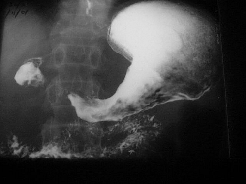
May be caused by Ulcer or carcinoma of the gastric antrum, Ulceration or scarring of the duodenal cap, Pancreatic carcinoma or duodenal carcinoma involving the duodenal loop or Infantile pyloric stenosis. The stomach is distended and often grossly enlarged with resting juice and food residue. A barium meal shows a mottled appearance of the barium as it mixes with the food residue and there is either no gastric emptying or marked delay. The cause of the obstruction is often difficult to demonstrate due to the large amount of food residue present in the stomach.
Infantile pyloric stenosis is now commonly diagnosed by ultrasound but if local expertise or the correct probe frequency is not available barium meal may be necessary.
Polyps
Polyps are relatively uncommon in the stomach compared to the large bowel. They are usually benign but if in the gastric antrum may be pre-malignant. Occasionally a leiomyoma is seen in the stomach. This is a benign tumour arising from the muscle layers. On barium meal, there is a smooth well- defined mass projecting into the stomach lumen. It may ulcerate with a central ulcer crater.
Lymphoma
May affect the stomach showing as very thickened folds or a large filling defect.
Duodenal deformity/ulceration
The most frequent site for a duodenal ulcer is the proximal part, the cap or bulb. Post bulbar ulcers may occur but are less common. Diagnosis depends on demonstration of a crater or niche, into which the barium pools. The crater may be anterior or posterior and chronic ulceration heals by scarring. This causes deformity of the cap, which often has a tri-lobed appearance if the ulcer crater is central. The scarring is permanent and reactivation of the ulcer, or ulcer healing is very difficult to detect on barium studies. Follow up of a duodenal ulcer is best done by endoscopy.
Use of Barium Meal in Diagnosis of Diseases affecting the small Intestine
Preferably the small bowel should be targeted for a single study with suitable barium mixture rather than done as a continuation of a barium meal (a follow through). The high density barium used for a satisfactory barium meal is not suitable for study of the small bowel where a larger volume of a relatively low density barium solution is more appropriate. It is better to do this as a separate study and the examination is now referred to as a small bowel meal. In some patients a small bowel enema may be necessary. In this examination, a tube is placed in the 3rd part of the duodenum and barium injected followed by air or water for double contrast. It shows the small bowel in greater detail but fluoroscopy is necessary.
Intestinal Obstruction
Occasionally a small bowel obstruction is not obvious on plain films. This is especially likely if the obstruction is very high or if the loops are filled with fluid rather than air. A small bowel study may localise the site and cause of an obstruction. Sometimes it is preferable to use water contrast such as gastrografin rather than barium. If surgery is needed immediately, it is easier if grossly distended loops of bowel are not full of barium, which will cause a serious peritonitis if spilled into the peritoneal cavity.
Malabsorption syndromes
These are best diagnosed clinically rather than by barium study although specific causes for the malabsorption may be demonstrated, such as Crohns disease. Coeliac disease causes non-specific dilatation of small bowel loops in severe cases but small bowel biopsy is much more specific. Jejunal diverticulosis, blind loops, fistulae and strictures may all cause malabsorption and are detectable on contrast studies.
Radiological features seen in malabsorption
- Dilation of the small bowel
- Thickening of the valvulae conniventes
- Clumping of the barium (flocculation) which does not maintain a continuous column.
Inflammatory bowel disease
Inflammatory diseases such as Crohns disease causes mucosal oedema with thickening of the folds, There may be strictures, and dilated loops especially in Crohns disease.
Lymphoma
Lymphoma may involve the small bowel. There is usually mucosal oedema together with displacement and distortion of bowel loops. Primary carcinoma of the small bowel is rare but can occur and usually presents as an obstruction.


