The gram stain procedure is a process that involves different steps, methods, or techniques. Gram staining results are used to differentiate bacteria as gram-positive or gram-negative. In microbiology, the gram stain procedure is one of the most crucial staining techniques.
This gram staining technique helps in the diagnosis of a disease or a pathologic condition. The staining technique was named after the Danish bacteriologist, Hans Christian Gram. Hans Gram, the danish bacteriologist was the first to introduce it in 1882, for the identification of pneumonia-causing organisms. This article is aimed at providing the step-by-step gram staining procedure explanation as well as the interpretation and meaning of the gram stain procedure results you’re likely to see.
Table of Contents
- What is a gram stain technique?
- Gram stain principle
- List of materials used in the gram staining procedure
- Steps of a gram stain procedure
- Gram stain procedure: gram staining steps and results
- List of the steps of gram staining procedure in order
- Gram stain results
- Recommendations
- FAQs on gram stain procedure
What is a gram stain technique?
Right from the first test performed, the gram staining technique involves the use of methylene blue or crystal violet as the primary stain. The interpretation of the gram stain results was based on the color seen after the sample organism is viewed under a microscope.
The organism that retains the primary stain in the Gram stain procedure and appears purple-brown under the microscope are termed Gram-positive organisms. Whereas the organisms termed gram-negative do not take up the primary stain and appeared red under the microscope. This staining technique helps in the differentiation and classification of microbes. Hence, the gram staining procedure is best described as a differential staining technique.
Importance and Application
As a differential staining technique, the gram staining procedure distinguishes bacterial cells based on the properties of their cell wall. There are two types of bacteria based on their cell wall composition- Gram-positive and gram-negative. About 90% of the gram-positive bacteria cell wall is made of thick layers of peptidoglycan whereas the gram-negative bacteria cell wall is composed of high lipid content as well as thin layers of peptidoglycan that only make up 10% of the cell wall.
As earlier said, the gram-positive bacterial cell walls have a thick layer of peptidoglycan, a protein-sugar complex, and low lipid content which is stained with the primary stain, and later fixed with a mordant. When this cell is decolorized, its thick cell wall is forced to get dehydrated and shrinks. This causes the pores in the cell wall to close and thus prevents the primary stain of the crystal violet dye from exiting the cell. Therefore, the decolorizer (ethanol solvent) cannot remove the crystal violet-iodine complex (CV-Iodine) that is bound to the thick layer of peptidoglycan of the bacterial cell wall. This is why the gram-positive bacteria appear blue or purple in color after staining.
Limitations of use
Even though the gram staining procedure is best described as a differential staining technique, it can’t be used for some organisms like the archaea or eukaryotes. The gram staining procedure and principle don’t work with these organisms because they lack peptidoglycan.
Gram stain principle
The principle of the gram stain involves the ability of the bacterial cell wall to retain the crystal violet dye during solvent treatment. Based on the cell wall of bacterial cells, the gram-positive microbes have higher peptidoglycan content, while the gram-negative microbes have higher lipid content. As the bacterial sample is stained with the primary stain crystal violet and fixed with the mordant-iodine, some of them are able to retain the primary stain while some are decolorized by alcohol.
All bacteria take up the crystal violet dye initially but when a solvent is used, the lipid layer from gram-negative microorganisms dissolves. As the lipid layer is dissolved, the gram-negative microorganism loses the primary stain. This is different for the gram-positive organisms because their cell wall is dehydrated by the solvent, because the closure of the pores of the gram-positive cell wall prevents the diffusion of the crystal violet-iodine complex and so the bacteria remain stained, making it gram-positive.
List of materials used in the gram staining procedure
Equipment
- Bunsen burner
- Alcohol-cleaned microscope slide
- Slide rack
- Microscope
Gram stain reagents
- Crystal violet as the primary stain
- Gram’s iodine solution as the mordant
- Acetone or ethanol as the decolorizer
- Safranin or 0.1% basic fuchsin solution as the counterstain
- Water
In the procedure of gram staining, Christain Gram initially used Gentian violet as the primary stain. However, crystal violet is mostly used today. There are recent modifications of the gram stain method published. The first one was by K. N. Atkins, who proposes the use of aniline sulfate in place of aniline, and a stronger iodine solution to which sodium hydroxide has been added.
The other modified gram stain method is by Burke which proposes the use of a 1% aqueous solution of dye, sodium bicarbonate, and acetone as a decolorizer. In Burke’s modification of the gram stain, sodium bicarbonate is added to the crystal violet solution. As iodine oxidizes, this sodium bicarbonate prevents the acidification of the crystal violet solution. An aqueous solution of safranin is used as the counterstain for this gram stain procedure modification.
Another modification method done by G. J. Hucker is also used today. Ammonium oxalate is added in Hucker’s method to prevent the precipitation of the dye. Also, an alcoholic solution of the counterstain is used in this method. Nevertheless, the above-listed reagents needed for the gram staining process can be purchased commercially or made.
Proper sequence of reagents in the gram stain procedure
The proper sequence of reagents in the gram stain procedure is:
- The crystal violet stain as the primary stain
- Then the mordant (gram iodine)
- The decolorizer
- Lastly the counterstain. The counterstain for the gram stain procedure can be safranin or basic fuchsin solution.
Crystal Violet Staining Reagent
In order to obtain the crystal violet staining reagent, the two solutions A and B are mixed together. After mixing, store for 24h and filter through filter paper before use.
Components of Solution A for crystal violet staining reagent
- 2g of Crystal violet (certified 90% dye content)
- 20ml of Ethanol, 95% (vol/vol)
Components of Solution B for crystal violet staining reagent
- 0.8 g of Ammonium oxalate
- 80ml of Distilled water
Gram’s Iodine
Components
- 1.0 g of Iodine
- 2.0g of Potassium iodide
- 300 ml of Distilled water
Formation
Grind the potassium iodide and iodine together. With continuous grinding add water slowly until the iodine is dissolved. Then store in amber bottles.
Decolorizing Agent
This is made up of:
- 50 ml Acetone
- 50 ml Ethanol (95%)
There are various formulations of decolorizing agents which include ethanol, acetone, and acetone/ethanol that are being used today. The most rapid decolorizer is acetone followed by acetone/ethanol, then ethanol. However, ethanol is recommended for student use in order to prevent over decolorization of sample cells.
Some professionals prefer using a 1:1 acetone and ethanol mixture whereas others use an acetone decolorizer. However, there is a variety of mixtures that are available commercially. Most commercial mixtures are 25 – 50% acetone with ethanol. A few add a small quantity of methanol or isopropyl alcohol in the formulation.
Counterstain – Safranin
You would need two types of solutions to form the counterstain reagent. These two solutions are the stock and working solutions.
Stock solution
The components of the stock solution are:
- 2.5g Safranin O
- 100 ml 95% Ethanol
Working solution
To form the working solution, combine 10 ml of the Stock Solution with 90 ml of distilled water.
Steps of a gram stain procedure
Gram stain procedure: gram staining steps and results
Equipment
- 1 Bunsen burner
- 2 Alcohol-cleaned microscope slide
- 1 Slide rack
- 1 Microscope
Materials
- 2 g Crystal violet
- 20 ml Ethanol
- 0.8 g Ammonium oxalate
- 80 ml Distilled water
Instructions
Application of the primary stain (crystal violet)
- The first of the gram stain steps is the application of the crystal violet (primary stain) to a heat-fixed smear. In this step, the crystal violet dye is used for the slide's initial staining.

Addition of a mordant (Gram's Iodine)
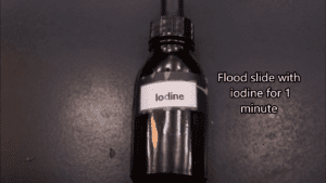
- The second step in gram staining is the addition of gram's iodine which is also known as fixing the dye. Iodine serves as a mordant in the gram staining procedure. This involves using iodine to form a crystal violet-iodine (CV-Iodine) complex to prevent easy removal of the crystal dye. Therefore, the role of gram iodine in the gram stain procedure is to fix the primary stain and prevent the crystal violet from leaving the cell.

Rapid decolorization
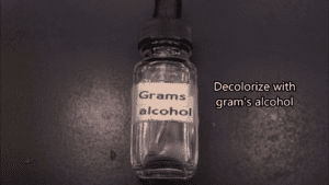
- Decolorization with alcohol, acetone, or a mixture of alcohol and acetone is the third phase of the steps in gram staining. This third step of decolorization is a very crucial step among the 4 steps of a gram stain. This is because prolonged exposure to a decolorizing agent can remove all the stains from both gram-positive and gram-negative bacteria. Solvents of ethanol and acetone are often used as decolorizers to remove the dye. The basic mechanism behind this particular gram stain process is the ability of the bacterial cell wall to retain the crystal violet dye during solvent treatment. Based on the cell wall structure of bacterial cells, the gram-positive microbes have higher peptidoglycan content, while the gram-negative microbes have higher lipid content. All bacteria take up the crystal violet dye initially but when a solvent is used, the lipid layer from gram-negative bacteria dissolves. As the lipid layer is dissolved, the gram-negative microorganism loses the primary stain. This is different for the gram-positive organisms because their cell wall is dehydrated by the solvent. The closure of the pores of the gram-positive cell wall prevents the diffusion of the violet-iodine complex and so the bacteria remain stained, making it gram-positive.

Counterstaining
- The fourth and final phase of the gram stain steps is to use a counterstain. Usually, the counterstain used in the gram staining procedure is Safranin or basic fuchsin stain. This process is known as counterstaining and the aim is to give the decolorized gram-negative bacteria pink color for easier identification.

Video
Notes
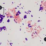
At the end of the gram staining procedure, gram-positive bacteria will be blue to purple in color whereas gram-negative bacteria will be red to pink in color.
Gram staining procedure in a flowchart
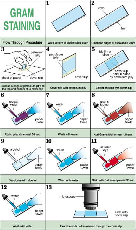
Photo Credit: www.cs.montana.edu
From the gram stain procedure explained, using the flowchart above as a quiz flashcard, you can mix up the images and then place the images in the correct order to assess your knowledge of the gram stain procedure.
What takes place during the gram stain process?
The crystal violet in an aqueous solution dissociates into CV+ and Cl– ions. These ions pass through the wall and membrane of both gram-negative and positive cells. The bacterial cells are stained purple as the CV+ interacts with negatively charged components of the bacterial cells. When the mordant is added, iodine (I– or I3) interacts with CV+. This forms large CV-I complexes within the cell’s cytoplasm and outer layers.
As a decolorizer is added, it interacts with the lipids of the membranes of both gram-positive and gram-negative cells. In gram-negative cells, the outer membrane of the cell is lost and the peptidoglycan layer is left exposed. There are just one to three thin layers of peptidoglycan in gram-negative cells which is a different structure from the peptidoglycan of gram-positive cells. The cell walls of gram-negative cells following ethanol treatment become leaky and allow the large CV-I complexes to be washed from the cell.
The principle behind the gram stain explains why the gram-negative bacteria doesn’t appear purple or blue in color as the gram-positive is associated with the thin layer of peptidoglycan. The gram-negative bacterial cell wall also takes up the crystal violet-Iodine (CV-Iodine) complex like the gram-positive bacteria. However, the CV-Iodine gets washed off due to the thin layer of peptidoglycan and the thick outer layer of lipids in the gram-negative bacterial cell wall. The lipids in the cell wall get dissolved when exposed to alcohol. The dissolution of the lipids due to the decolorizer allows the CV-Iodine complex to leach out of the cells. This is the reason why when these cells are then stained with safranin, they take the stain color and appear red in color.
After the addition of ethanol, the highly cross-linked and multi-layered peptidoglycan of the gram-positive cell is dehydrated. Due to dehydration from the solvent treatment, the multi-layered nature of the peptidoglycan traps the large CV-I complexes within the cell. The gram-positive cell remains purple in color after decolorization whereas the gram-negative cell loses the purple color. The loss of the purple color in the gram-negative cell is only revealed when the positively charged counterstain, safranin is added.
List of the steps of gram staining procedure in order
- Make a smear of the culture cells that is not too heavy or too light on a slide.
- Air-dry the slide and then move it over the flame to be heat-fixed.
- Flood the heat-fixed smear of cells with crystal violet staining reagent for 1 minute.
- After flooding, rinse the slide under a gentle and indirect stream of water for at least 2 seconds.
- Then, flood the slide with gram’s iodine and wait for 1 minute.
- Rinse the slide again under a gentle and indirect stream of water for at least 2 seconds.
- Then, flood the slide with a decolorizing agent and rinse with water in 5 seconds.
- Counterstain by flooding the slide with safranin, then wait for 30-60 seconds before rinsing.
- Rinse the slide under a gentle and indirect stream of water until no color is seen in the effluent.
- Blot dry slide with absorbent paper and then view the slide under oil immersion.
- Use a brightfield microscope to view the specimen.
- After gram staining, gram-negative bacteria will stain pink or red while gram-positive bacteria will stain purple or blue.
Explanation on the gram stain procedure steps
It is important to know all parts of the gram stain procedure and the rationale of each step. There are time durations and precautions for each step that needs to be adhered to. Below is a detailed gram staining procedure explanation and precautionary measures.
Prepare a slide smear
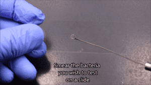
The first process in the procedure of gram staining is to prepare a slide smear of the cell. A few loopful or a drop of water is added to a petri dish or slant culture tube that has the colony. This is done to ensure a minimal amount of colony is transferred to the examination slide. Use an inoculation loop to transfer a drop of suspended culture to the microscope examination slide.
You only need a minimal amount of culture on your slide, if you can visually detect the culture on an inoculation loop, it means you have collected too much culture. It is important to note in the gram stain procedure, that, the quality of the smear will affect the gram stain results. The concentration of the smear culture should not be too heavy or too light.
With the inoculation loop, spread the culture to an even thin film over a circle of 15mm in diameter. In examining more than one culture, a typical slide can contain up to 4 small smears. After making your smear on the slide, you can air-dry the slide or run it over a gentle flame to dry.
Fix the slide
Move the slide circularly over the flame to avoid overheating or the formation of ring patterns in the slide. In the gram stain procedure, fixation is needed because running the slide over the flame enhances the cell adhesion to the slide and prevents significant loss of culture when you later rinse the slide after staining.
Gram stain the heat-fixed culture
Flood the fixed culture with crystal violet stain and after 1 minute pour the stain off and rinse the excess stain on the slide with water. Rinse gently, as the aim is to wash off the stain without losing the fixed culture. In the gram staining procedure, water serves as the neutral reagent for rinsing off the applied reagent.
After that, flood the smear again for 10 to 60 seconds with iodine solution. This particular gram stain process is known as fixing the dye. In the gram stain procedure, iodine serves as a mordant that fixes the crystal violet to the bacterial cell wall. Pour the iodine solution off and then rinse the slide with running water.
Shake off the excess water from the surface of the smear, then add a few drops of decolorizer to the slide. This particular procedure of gram staining is known as a solvent treatment and is the most crucial step of all the gram stain procedure steps.
In 5 seconds, ensure you rinse the slide with water. The decolorizers are usually mixed solvents of ethanol and acetone and excess decolorization is not appropriate. In fact, it is said that the step in the gram stain procedure that is most prone to error is the decolorization step.
It’s advisable to stop adding the decolorizer as soon as the solvent flows over the slide giving a colorless effluent. After you have rinsed the decolorizer off the slide, stain the smear with a basic fuchsin solution or safranin. This gram stain process is known as counterstaining. The counterstain in a gram stain procedure is the basic fuchsin solution or safranin.
After staining with the counterstain, wait for 30-60 secs and then wash off with water and with bibulous paper blot the excess water or air dry the slide after shaking off the excess water. As seen from the gram stain procedure explained, the proper sequence of reagents in the gram stain procedure is the crystal violet stain as the primary stain followed by the mordant (gram iodine), then the decolorizer, and lastly the counterstain (safranin or basic fuchsin solution).
The color of gram-positive bacteria after adding the decolorizing agent in the gram stain procedure remains purple in color, whereas the purple color of the crystal dye in gram-negative bacteria is lost after adding the decolorizing agent. This gram-negative bacteria is only revealed when the counterstain, the positively charged dye safranin, is added.
Examine the slide under a microscope
Examine the slide with a microscope under oil immersion. Use the X40 objective for the initial slide examination to evaluate the smear distribution. Then, the slide should be examined using the X1000 oil immersion objective.
It is important to note that all areas of the slide need an initial examination. Areas that are only one cell thick should be examined and thick areas in slides usually give variable and incorrect results. Use brightfield microscopy when viewing the slide and adjust the brightness sufficiently enough to reveal the color of the stained sample bacteria.
White blood cells and macrophages stain gram-negative while squamous epithelial cells stain gram-positive. Examples of gram-positive organisms include Staphylococcus species, Clostridium species, Streptococcus species, Corynebacterium species, and Listeria species. Gram-negative organisms examples include Pseudomonas species, Neisseria meningitidis, Proteus species, Moraxella species, Neisseria gonorrheae, Escherichia coil, and Klebsiella species. Actinomyces species are examples of gram variable organisms.
Gram stain procedure video
Below is a gram stain procedure video showing how the gram staining reagents and other materials are used appropriately during the gram stain process.
At the completion of the procedure of the gram stain, the gram-positive cell is purple or blue and the gram-negative cell is pink to red. However, after staining with the gram Stain, some bacteria yield a pattern called gram-variable where a mix of pink and purple cells are seen. There are some bacteria genera with cell walls that are specifically sensitive to breakage during cell division e.g Mycobacterium, Actinomyces, Corynebacterium, Arthrobacter, and Propionibacterium. This breakage results in these gram-positive cells staining as gram-negative cells.
Using the gram stain, some bacteria do not stain as expected. Typical examples include:
- Bacteria of the genus Acinetobacter that are usually resistant to the decolorization step. After a well prepared gram stain, these gram-negative cocci usually appear gram-positive.
- Even though Mycobacteriym spp is considered to be gram positive, the waxy nature of the coat of this bacterium makes itnot readily stainable with the dyes used in the Gram stain process.
- Bacteria of the genus, Gardnella possess an unusual gram-positive cell wall structure that causes them to stain gram-negative or gram-variable.
Additionally, the age of the culture may interfere with the results of the stain in all bacteria stained using the gram stain. Misinterpreting the gram stain has led to delayed diagnosis or misdiagnosis of infectious disease.
Gram stain results
The gram stain procedure’s purpose is to differentiate bacterial cells into gram-positive or gram-negative cells. Hence, it is important to arrange the steps of the gram staining procedure in their correct order. Do not overlap any steps as each of the techniques used in the process is aimed at getting the right gram stain results. Gram-negative bacteria cells will become red/pink at the end of a gram stain procedure while gram-positive bacteria cells will become purple/blue.
Whenever a bacterial infection is suspected, gram staining is indicated for easy and early diagnosis. After microscopic examinations, the finding is said to be normal, if there are no pathologic organisms found in the smear. However, if organisms are found, they are identified in the smear based on color and shape.
Points to note for interpretation of the gram stain results
In order to interpret your gram stain results it is important to note the following:
- When the organism identified is either purple or blue in color, it is gram-positive.
- An organism that appears either pink or red in color is gram-negative.
- There are gram-variable organisms that do not group into either gram-positive or gram-negative.
- Bacilli is a class of bacteria that are rod-shaped.
- Cocci is a class of bacteria that are spherical.
- There are some bacteria that are shaped like very short rods or ovals known as “Coccobacilli”. This name was derived from combining the words “cocci” and “bacilli”.
- Diplococci are bacteria that occur as pairs of cocci.
Study the gram stain results table and interpretations below to be able to understand the findings you’re likely to get during microscopic examination of the smear after staining. This table will help you interpret the diagnostic information from the gram stain procedure.
Gram staining results (Findings on gram stain) | Interpretations | |
1 | Gram-positive cocci in clusters | This suggests the presence of Staphylococcus species such as S. aureus |
2 | Gram-positive cocci in chains | This can indicate the presence of Streptococcus species such as S. pneumoniae, B group streptococci |
3 | Gram-positive cocci in tetrads | This suggests the presence of Micrococcus spp. |
4 | Gram-positive bacilli and thick | This is a characteristic of Clostridium spp. and suggests the presence of species such as C. perfringes, C. septicum |
5 | Gram-positive bacilli and thin | This suggests the presence of Listeria spp. |
6 | Gram-positive bacilli branched | This is usually characteristic of Actinomyces and Nocardia |
7 | Gram negative coccobacilli | This indicates the presence of Acinetobacter spp. Note: These organisms can be gram-positive, gram-negative, or gram variable. They can also appear as gram-positive cocci and can be pleomorphic. |
8 | Curved | This indicates the presence of Vibrio spp. or Campylobacter spp., such as V. cholerae, and C. jejuni. |
9 | Gram-negative bacilli and thin | This is a typical characteristic of Enterobacteriaceae, such as E. coli. |
10 | Gram-negative coccobacilli or bacilli, and small with random arrangements | This indicates the presence of Hemophilus spp., such as H. influenzae. |
11 | Gram-negative diplococci with flattened shape | This suggests the presence of Neisseria spp. such as N. meningitidis. Note: Moraxella spp is often diplococci in morphology and gram-negative resembling Neisseria spp. |
12 | Thin needle shape | This is a typical characteristic of Fusobacterium spp. |
Recommendations
There are avoidable reasons why a gram stain procedure may produce an incorrect result. For instance, if the microscopic smear is thick and clumped, interpreting the slide can be difficult. Ensure the smear of cells is not crowded because this will make it difficult for the cell shape and arrangement to be seen. View the gram-stained bacteria with a bright field microscope with oil immersion at X1000 magnification.
Also, it is highly recommended to use freshly made staining reagents but if using older staining reagents ensure you filter the staining reagents before use. Another important step that affects the gram stain procedure results is the decolorization step. Of all the gram stain procedure steps, decolorization is very crucial. The time of decolorization should be monitored closely to avoid under-decolorization or over-decolorization. Smears that are thicker tend to need longer decolorizing time.
Also, cultures should be evaluated while they are still fresh. Young and actively growing cultures are best used for gram staining. This is because old cultures tend to lose the peptidoglycan cell walls which will cause erroneous results. Losing the peptidoglycan cell wall will cause gram-positive cells to appear gram-negative or gram variable.
Moreso, an intact cell wall is needed for accurate gram stain results. Older cultures tend to have breaks in the cell wall which usually give gram variable results where pink or red cells are seen among blue or purple cells. Gram stain is not used for organisms that lack cell walls such as Mycoplasma species and smaller bacteria such as Rickettsia and Chlamydia species.
Also, it is recommended to use a gram stain control. Include a sample of cells with a known gram stain reaction on the same slide with the test culture. This will serve as a control for success in the gram stain technique. Furthermore, a KOH string test can be used as a confirmatory test for the gram stain. In 3% KOH, the formation of a string (DNA) within 60 secs suggests that the test organism is a gram-negative organism. In the KOH, gram-positive cells are usually not affected.
When counterstaining, the positively charged dye added replaces the crystal violet dye in the gram-positive bacteria and stains the gram-negative bacteria. Even though the mordant that was used slows down this process, it is still advisable and ideal to not overexpose cells to the counterstain. Counterstaining should not take more than 30-60 secs.
FAQs on gram stain procedure
What is the most critical step in the gram staining procedure
The most critical step in the gram staining procedure is the decolorizing step as well as the thickness of the smear. How thick the smear is will definitely affect the result of the gram stain and the decolorizing step affects the outcome of the stain.
Over-decolorizing will cause an erroneous result where gram-positive cells may appear pink or red suggesting a gram-negative result. Under-decolorizing on the other hand causes an erroneous result whereby gram-negative cells may appear purple or blue suggesting a gram-positive result.
Why is gram staining important?
Gram staining is important for the differentiation of bacterial organisms according to the structure of their cell wall. Hence, this differential staining technique is crucial to the phenotypic characterization of bacteria.
Bacterial cells that are gram-positive have a thick peptidoglycan layer and stain blue to purple while bacterial cells that are gram-negative have a very thin peptidoglycan layer and stain red to pink. Therefore, the gram stain procedure’s purpose is to differentiate bacterial cells.
Why is the gram staining procedure not appropriate for staining mycobacteria?
The gram staining procedure is not appropriate for staining mycobacteria, but the acid-fast stain is appropriate for these organisms. Why the gram staining procedure is not appropriate for staining mycobacteria is because mycobacteria as acid-fast bacteria have a high content of mycolic acids in their cell wall. Hence their cell wall tends to have characteristics of both gram-positive and gram-negative bacteria.
The thickness of their cell wall together with the mycolic acids makes these organisms not respond well to staining agents. For these bacteria, acid-fast stains are used to differentiate them with a staining technique called Ziehl–Neelsen staining. This bacteriological stain was first introduced by Paul Ehrlich. In this staining technique, acid-fast bacteria like the mycobacteria will stain red whereas the nonacid fast bacteria will stain blue or green.
Which step in the gram stain procedure is most prone to error if done incorrectly?
The decolorization step is more prone to error because the degree of decolorizing needed is determined by the thickness of the smear on the slide. It is recommended that the cells be prepared with a thin smear ensuring there are no areas of clumping or inconsistency. Also, for correct results, it is recommended that when staining the thin smear, one should use a short decolorizing time.
Some individuals recommend flooding the slide for 15 seconds or less with the decolorizing agent, whereas others recommend adding the decolorizing agent in drops for 5- 15 seconds or adding in drops until the color of the decolorizing agent running from the slide no longer show any color.
What would you expect to see if you forgot to perform step 2 of the gram stain procedure?
If you forgot to perform step 2 of the gram stain procedure, expect to see all bacteria cells appearing pink. This is because forgetting mordant in the gram stain procedure will not help fix the crystal dye and without iodine, the bacterial cells cannot retain the primary stain.
What is the function of a mordant and which reagent serves this purpose in the gram stain procedure?
The function of a mordant is to prevent the primary stain which is the crystal violet from leaving the cell. In the gram stain procedure, iodine serves as a mordant that fixes the crystal violet to the bacterial cell wall. It binds to the crystal violet and forms a large complex that adheres to the cell membrane.
What role does safranin play in the gram stain procedure?
The role safranin play is to act as a counterstain. In fact, the main purpose that safranin serves in the gram stain procedure is to differentiate the bacterial cells after decolorization. Safranin as a counterstain doesn’t disrupt the purple coloration in gram-positive cells because it is lighter than crystal violet. However, it stains the decolorized gram-negative cells red or pink.
What are the differences between simple staining and the gram stain procedure?
Simple staining involves the use of only 1 dye whereas the gram stain procedure as a differential staining technique involves the use of more than 1 dye.
Under what conditions might a gram-positive bacterium stain red following the gram stain procedure?
Over-decolorizing can cause a gram-positive bacterium to appear pink or red which suggests a gram-negative result.
What is the purpose of using methyl alcohol in the gram stain procedure?
The purpose of using methyl alcohol (Methanol) in the gram stain procedure is to fix the smear to the slide. This is an alternative and better fixation method compared to heat fixing. In order to fix the slide after making the smear, the slide is flooded with methyl alcohol for 1 minute. This will prevent the liquid specimen from washing off the slide. It also preserves the morphology of the cell and results in a clearer background.
What happens if you over-decolorize in the gram stain procedure?
Over-decolorizing will cause an erroneous result where gram-positive cells may appear pink or red suggesting a gram-negative result.
Why are we fixing the smears before applying the gram stain procedure?
Fixing the smears before applying the gram stain procedure is necessary because fixation enhances the cell adhesion to the slide and prevents significant loss of culture when you later rinse the slide after staining. Fixation can be done by using methanol or by heat fixing where you move the slide in a circular motion over the flame.
What are two different types of stains used for the gram stain procedure?
There are 2 types of stains used in a gram stain procedure; the primary stain and the counterstain. The primary stain in the gram staining procedure is crystal violet reagent while the counterstain is safranin.
If you leave the decolorizer for one minute in the gram stain procedure, what would be the result?
If you leave the decolorizer for one minute in the gram stain procedure, you would get an erroneous result. It is recommended that you decolorize between 5- 15 seconds, as anything more than that is over decolorization. Over-decolorization makes gram-positive cells appear pink or red which suggests a gram-negative result.
In the gram stain procedure, what is the role of alcohol?
The role of alcohol in the gram stain procedure is to serve as a decolorizer that dehydrates the peptidoglycan layer which makes the layer shrink and tighten up.
As a result, ethanol does not decolorize gram-positive cells in the gram stain procedure because the large crystal violet-iodine (CV-Iodine) complex finds it hard to penetrate this tightened peptidoglycan layer. Thus, the CV-Iodine complex is trapped in the gram-positive bacterial cell.
What is the percentage of alcohol used in the gram stain procedure?
95% Ethyl Alcohol is the recommended percentage of alcohol used in the gram staining procedure.
Could other dyes be substituted for safranin in the gram stain procedure?
Yes, Basic fuchsin stain can be substituted for Safranin in the counterstaining procedure. Even though many laboratories use safranin as a counterstain, basic fuchsin stains gram-negative organisms more intensely than safranin. Moreso, some bacterial species such as Hemophilus spp., Legionella app, and some anaerobic bacteria stain poorly with safranin.
What is the primary dye in the gram stain procedure?
In the gram stain procedure, crystal violet stain is the primary stain (dye).
What is the differential step in the gram stain procedure?
The decolorization step of the gram stain is the differential step. This is because the 95% ethanol removes the primary stain (crystal violet) from gram-negative cells. The bacteria would remain purple and give a positive result that is false if this step is missed.
What molecule is unique to bacterial cell walls and is the basis of the gram stain procedure?
The molecule unique to bacterial cell walls is the peptidoglycan. This molecule is the basis of the gram stain procedure because the bacteria are differentiated in this process based on their cell wall composition. About 90% of the gram-positive bacteria cell wall is made of thick layers of peptidoglycan whereas the gram-negative bacteria cell wall is composed of high lipid content as well as thin layers of peptidoglycan that only make up 10% of the cell wall.
The gram stain principle involves the ability of the bacterial cell wall to retain the crystal violet dye during solvent treatment. Based on the cell wall of bacterial cells, the gram-positive microbes have higher peptidoglycan content, while the gram-negative microbes have higher lipid content. As the bacterial sample is stained with the primary stain crystal violet and fixed with the mordant-iodine, the gram-positive cells are able to retain the primary stain while the gram-negative cells are decolorized by alcohol.
What part of the bacteria does the gram staining procedure stain?
The cell wall of the bacteria.
This staining procedure mechanism involves the ability of the bacterial cell wall to retain the crystal violet dye during solvent treatment. According to the cell wall composition of bacterial cells, the gram-positive cell wall has higher peptidoglycan content, while the gram-negative cell walls have higher lipid content.
As the bacterial sample is stained with the primary stain, crystal violet, and fixed with the mordant-iodine, the positive cell wall retains the primary stain after decolorization while the negative cell walls are decolorized by alcohol.
What color is a “gram-positive” bacterium after completion of the gram staining procedure?
Blue or purple is the color of gram-positive bacteria after the completion of the staining procedure.
What are the initial and secondary stains used in a gram staining procedure?
The initial stain used is the crystal violet regent which serves as the primary stain then the secondary stain used after decolorization is safranin which serves as the counterstain. This simply means the basic stains used in the gram staining procedure include the primary stain and the counterstain.
What is the correct order of the reagents used in the gram staining procedure?
The correct order of the reagents used in the procedure of gram stain is the crystal violet stain, the mordant (gram iodine), decolorizing agent, and lastly the counterstain (safranin).
In the gram stain procedure, which phase of the stain is considered the mordant?
The second phase of the gram stain process which involves the addition of the grams iodine is considered the mordant.
Can you use methylene blue as a counterstain in the gram stain procedure?
Methylene blue would not give as strong of a contrast to the crystal violet the way the safranin does.
What color would be seen if the alcohol were left out of the gram stain procedure?
If you don’t use alcohol during the gram stain procedure, the gram-negative bacteria will retain their outer cell membrane and stay purple when the staining is done. This means both gram-positive and negative bacteria will stain purple if alcohol is left out of the gram staining process.
Gram-negative bacteria will be stained what color at the end of the gram staining procedure?
Gram-negative bacteria will become red/pink in color at the end of the procedure while gram-positive bacteria will be stained blue to purple in color.
What is the mordant in the gram stain procedure?
The mordant used in the Gram stain procedure is iodine. As a mordant, the role of gram’s iodine in the procedure of gram staining is to promote dye retention.
What are the steps of a gram stain in order?
The order of steps in the gram staining procedure is the application of primary stain, addition of the mordant, rapid decolorization, and counterstaining.
