Table of Contents
Do bacteria have cell walls?

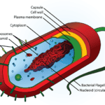
Bacteria do have a cell wall, though not all of them. The bacterial cell is enclosed in a cell envelope that consists of the cell membrane and cell wall. As in the plant cell, the bacterial cell wall structure gives the cell structural integrity. The main function of the cell wall in prokaryotes is to give the cell protection. Compared to the external environment of the cell, there are higher concentrations of proteins and other molecules in the cell. Hence, the cell wall protects the cell from internal turgor pressure that such high concentrations can cause.
Nevertheless, in microbiology the cell wall of bacteria is not like that of plants and other organisms due to the presence of peptidoglycan (also known as murein) which is located immediately outside the cell membrane.
The bacteria cell wall is made up of peptidoglycan which makes the cell wall rigid and determines the cell shape. The structure of peptidoglycan is relatively porous and is not considered a permeable barrier for small substrates. It consists of a polysaccharide structure that has an equal amount of alternating N-Acetylmuramic acid (NAM) and N-acetylglucosamine (NAG) residues.

Why do bacteria have cell walls?
The cell wall is needed for bacteria survival and protection from being detected by antibodies; this is why antibiotics are used to treat bacterial infection because the cell wall is able to evade being detected by the body’s immune system but antibiotics can cause pores in the cell wall of bacteria, thereby denaturing and destroying the bacteria. Several antibiotics such as cephalosporins and penicillins interfere with cell wall synthesis in order to stop bacterial infection. These antibiotics have no effects on human cells because animal cells don’t have a cell wall (only a cell membrane).
In the bacterial cell wall, beta-lactam antibiotics like penicillin restrict the formation of peptidoglycan cross-links. Also, the enzyme lysozyme found in the tears of humans, serving as a defense against eye infections breaks down the bacterial cell wall. Therefore, it is called a protoplast once the cell wall of the bacterium is removed entirely. If the cell wall of the bacteria is removed partially, it is called a spheroplast.
As the cell keeps moving from one environment to another, water can freely move from the cell membrane and cell wall making the cell at risk of osmotic imbalance leading to osmotic lysis of the cell. Due to this, an important function of the bacterial cell wall is to protect the cell from osmotic lysis. The bacterial cell wall function in keeping certain toxic molecules out of the cell. It also plays a role in the pathogenicity (disease-causing ability) of bacterial cells.
The majority of bacterial cells have a cell wall and there are two types of cell walls. Bacteria could either have a gram-positive cell wall or a gram-negative cell wall. These two types of bacterial cell walls are differentiated by their Gram staining characteristics. Particles of approximately 2 nm can pass through the peptidoglycan of both the gram-positive and gram-negative bacterial cell walls.
Bacteria without a cell wall
The bacteria that lack a cell wall belong to the phylum Tenericutes. There is a group of them called mycoplasmas. Due to the absence of a cell wall, they are very susceptible to osmotic changes. Hence, they usually inhabit osmotically-protected environments. In order to strengthen their cell membrane, sterol-like molecules are incorporated into their membranes. These sterols are normally associated with the cell membranes of eukaryotes.
The majority of bacteria that belong to the phylum Tenericutes choose to hide out as pathogens in the protective environment of a host. For example, Mycoplasma pneumonia causes bacterial pneumonia also known as walking pneumonia and for some reason, penicillin can’t treat this kind of pneumonia.
Pathogenic bacteria can sometimes revert to cell wall-less forms (protoplasts or spheroplasts) when under the pressure of antibiotic therapy. In this cell wall-less form they persist and survive in osmotically protected tissues. These bacteria may regrow their cell walls when the antibiotics are withdrawn. Then, reinfect unprotected tissues of the host.
Types of bacterial cell wall
- Gram-positive cell wall
- Gram-negative cell wall
The two main types of cell walls can be identified in the lab using a differential stain called the Gram stain which has been in use since it was developed in 1884. Initially, it was not known why the gram stain could reliably separate bacteria into these two groups. However, it was when the electron microscope was invented that it was discovered that the staining difference was correlated with differences in the cell walls of bacteria. The gram stain when applied will stain the gram-positive bacteria purple and stain the gram-negative bacteria pink.
Gram-positive cell wall
This cell wall type is thick and gram-positive bacteria take up the crystal violet dye of the stain and thus are stained purple. In some gram-positive bacteria, the structure of the peptidoglycan layer makes up 95% of the gram-positive cell wall. Peptidoglycan consists of a polysaccharide structure that has an equal amount of alternating N-Acetylmuramic acid (NAM) and N-acetylglucosamine (NAG) residues.
In the gram-positive bacterial cell wall structure, the matrix substance may be polysaccharides or teichoic acids. Teichioc acids are very widespread, though they are only found in gram-positive bacteria. These teichoic acids are of two main polymer types which include ribitol teichoic acids and glycerol teichoic acids. Glycerol teichoic acids tend to be more widespread among the two types.
Ribitol teichoic acids are polymers of ribitol phosphate while glycerol teichoic acids are polymers of glycerol phosphate. These acids are only located on the surface of several gram-positive bacteria. Lipoteichoic acid which renders an antigenic function is the main component of the gram-positive cell wall. The lipid element is seen in the cell membrane and its adhesive properties help it to anchor to the membrane.
Lysozymes can completely dissolve the cell wall of some gram-positive bacteria. The bonds between N-acetylmuramic acid and N-acetylglucosamine are attacked by this lysozyme enzyme. However, other gram-positive cell walls e.g Staphylococcus aureus are resistant to lysozymes. This is because they have O-acetyl groups on carbon-6 of some muramic acid residues.
Examples of bacteria with gram-positive cell wall
- Micrococcus
- Staphylococcus
- Streptococcus
- Leuconostoc
Gram-negative cell wall
This cell wall type is much thinner compared to the gram-positive cell walls. In gram-negative bacteria, the peptidoglycan layer constitutes as little as 5-10% of the cell wall. The gram-negative cell wall has a second plasma membrane that is superficial to the peptidoglycan layer and adjacent to the cytoplasmic membrane. This cell wall type is more complex than the gram-positive cell wall and the gram staining technique stains the gram-negative bacteria pink.
The gram-negative cell wall has a plasma membrane called the outer membrane. This membrane is located outside of the peptidoglycan layers and is composed of a lipid bilayer. The outer membrane of the cell wall has large molecules known as lipopolysaccharides (LPS). These LPS are anchored into the outer membrane and project into the environment from the cell. The chemical structure of the LPS is usually distinct to specific bacterial subspecies. It plays a role in many of the antigenic properties of these strains of bacterial subspecies.
Lipopolysaccharides consist of three different components which include:
- O-antigen or O-polysaccharide (the outermost part of the structure)
- The core polysaccharide
- Lipid A that anchors the LPS into the outer membrane
Examples of bacteria with gram-negative cell wall
- E. coli
- Salmonella
- Klebsiella
- Shigella
- Pseudomonas
Gram-positive cell wall vs Gram-negative cell wall
Gram-positive cell wall | Gram-negative cell wall | |
Cell wall thickness | 15-80nm | 2nm |
Teichoic acid | Present | Absent |
Amino acid variety | Few | Many |
Sulfur-containing amino acids | Absent | Present |
Lipid content | 2-5% | 15-20% |
Aromatic amino acids | Absent | Present |
Treatment with lysozyme | Protoplast | Spheroplast |
Bacterial cell wall structure
The cell wall is located outside the cell membrane and serves as an additional layer that gives the strength that the cell membrane lacks. The bacterial cell wall structure is semi-rigid and contains an element known as peptidoglycan. This particular substance which is also called murein is not seen anywhere else other than the cell wall of bacteria.
There are other additional components that make up the bacterial cell wall, which gives it an overall complex structure as compared to the cell wall of eukaryotes. The cell walls of eukaryotic microbes are made up of a single component e.g chitin in fungal cell walls and cellulose in the algal cell walls. Whereas in the bacterial cell wall, three other main components are present outside the peptidoglycan layer. These components include the lipoprotein layer, outer membrane, and lipopolysaccharides. The gram-positive bacteria cell wall is made up of peptidoglycan, lipid, teichuronic acid, and teichoic acid. Whereas, the gram-negative bacteria cell wall is made up of peptidoglycan, and the outer membrane (that consists of lipid, protein, and lipopolysaccharides).
Structural components
- Peptidoglycan
- Teichoic acid
- Teichuronic acids
- Outer membrane
- Lipopolysaccharides LPS
- Lipoprotein layers
Peptidoglycan
This substance is also called murein and is common in the two types of bacterial cell walls. The structure of peptidoglycan is a polysaccharide that is composed of two glucose derivatives that are alternated in long chains. These glucose derivatives include N-acetylglucosamine (NAG) and N-acetylmuramic acid (NAM). A tetrapeptide extending off the NAM sugar unit cross-links the chains to one another forming a lattice-like structure. This tetrapeptide is made up of 4 amino acids, namely:
- L-alanine
- D-glutamine
- L-lysine or Meso-diaminopimelic acid (DPA)
- D-alanine
Cells utilize only the L-isomeric form of amino acids. However, utilizing the mirror image D-amino acid gives protection from proteases that attack the peptidoglycan and might compromise the cell wall. The tetrapeptides can be cross-linked directly to one another with the L-lysine/DPA on one tetrapeptide binding to the D-alanine on another tetrapeptide.
There is a cross-bridge of 5 amino acids like glycine (peptide interbridge) in many gram-positive bacteria that connect one tetrapeptide to a different one. The cross-linking in whatever case serves to extend the general strength of the structure. More strength is therefore derived from complete cross-linking as every tetrapeptide is bound one way or the other to a tetrapeptide on another NAG-NAM chain. In other words, peptidoglycan is claimed to be synthesized as a cylinder with a coiled substructure that has each coil cross-linked to subsequent coil forming a stronger overall structure.
Even though peptidoglycan is an important component to both the gram-positive and gram-negative bacteria, there are some exceptions. For instance, bacteria that belong to the phylum Chlamydiae, in all other regards (LPS, porin, outer membrane, etc) tend to have a gram-negative cell wall structure but lack peptidoglycan. It is said that these bacteria may be using a protein layer that functions the same way the peptidoglycan does. This is likely of advantage to the cell as it provides resistance to β-lactam antibiotics (e.g penicillin) that attack peptidoglycan.
Teichoic acid
This is a water-soluble polymer of polyglycerol phosphate or ribitol phosphate that is present in gram-positive bacteria. A covalent bond connects these polymers to the peptidoglycan. Teichioc acids may have amino acid or sugar substitutes within the chains or as side chains. As a major surface antigen, it makes up about 50% of the dry weight of the gram-positive cell wall. These polymers are involved in the regulation of cell morphology and cell division.
Teichioc acids are very widespread, though they are only found in gram-positive bacteria. These teichoic acids are of two main polymers which include ribitol teichoic acids and glycerol teichoic acids. Glycerol teichoic acids are more widespread among the two polymers. Ribitol teichoic acids are polymers of ribitol phosphate while glycerol teichoic acids are polymers of glycerol phosphate. These acids are only located on the surface of several gram-positive bacteria.
There are two categories of teichoic acids:
- Wall teichoic acid (WTA)
- Lipoteichoic acid or Lipid teichoic acid(LTA)
The WTA and LTA are covalently bound to the peptidoglycan or embedded into the cell membrane. Lipoteichoic acid and wall teichoic acid have different metabolism pathways even though they share common composition. They are involved in the control of cell division and cell morphology. Also, WTA and LTA contribute to cellular adhesion. Lipid teichoic acids render an antigenic function as the main component of the gram-positive cell wall. The lipid element is seen in the cell membrane and its adhesive properties help it to anchor to the membrane.
Teichuronic acids
This teichuronic acid is made up of repeated units of sugar acids. They are gram-positive bacterial cell wall components that represent phosphate-free and uronic acid-containing polysaccharides. These acids are normally produced in small quantities and only when there is phosphate deficiency.
When the phosphate supply in the cell is not sufficient enough, the teichuronic acids are synthesized as substitutes for teichoic acids. Phosphate deficiency results in a stop in the biosynthesis of teichoic acids. These teichuronic acids are produced in higher quantities and assume control of the teichoic acid function e.g binding of metal ions.
Outer membrane
This is an additional layer in gram-negative bacteria that comprises a lipid bilayer, a protein, and a lipopolysaccharide (LPS) layer. The outer membrane is described as a discrete bilayered structure that is present outside of the peptidoglycan sheet. It superficially resembles the plasma membrane as it is a lipid bilayer that is intercalated with proteins. This membrane is located outside of the peptidoglycan layers.
Similar to the phosphoglycerides that make up the plasma membrane, the phospholipids make up the inner face of the outer membrane. The outer face of the outer membrane is mainly formed by a different kind of amphiphilic molecule that is made up of lipopolysaccharides (LPS). However, the outer face may still contain some phospholipid that makes up the inner face of the outer membrane.
Therefore, the inner part of the outer membrane resembles the plasma membrane in composition whereas the outer face is distinct. Usually, the outer membrane proteins transverse the membrane, anchoring the outer membrane to the underlying peptidoglycan sheet. The outer membrane proteins and porins are the proteins in the outer membrane.
Lipopolysaccharides (LPS)
LPS is made up of lipid-A and polysaccharides. The lipid-A is a phosphorylated glucosamine disaccharide that is antigenic and the polysaccharide consists of core-polysaccharide and O-polysaccharide. There is a hydrophobic bond that attaches the LPS to the outer membrane. The lipopolysaccharide is synthesized in the cytoplasmic membrane and sent to the outer membrane.
These LPS are anchored into the outer membrane and project into the environment from the cell. The chemical structure of the LPS is usually distinct to specific bacterial subspecies. It plays a role in many of the antigenic properties of these strains of bacterial subspecies.
The LPS is an endotoxin that is produced by gram-negative bacteria. LPS are actually complex molecules that are adhesive in nature as they help the gram-negative bacteria to adhere. The LPS serves several functions for the cell such as:
- It contributes to the net negative charge for the cell.
- LPS helps to stabilize the outer membrane.
- As the LPS blocks access to other parts of the cell wall, it provides protection from certain chemical substances.
- It plays a role in the host response to pathogenic gram-negative bacteria.
In an infected host, the O-antigen triggers an immune response that causes the production of antibodies specific to that part of LPS. Lipid A serves as a toxin (endotoxin), that causes general symptoms of illness such as diarrhea and fever. An endotoxic shock can be triggered when a large amount of lipid A is released into the bloodstream. Endotoxic shock is a body-wide inflammatory response that can be life-threatening.
Lipoprotein layers
The lipoprotein layer is composed mainly of special lipoproteins known as Braun’s lipoproteins. These lipoproteins that are covalently joined to the underlying peptidoglycan layer are very small. With their hydrophobic end, they are incorporated into the outer membrane.
Functions of bacterial cell wall
- A major function of the bacterial cell wall is that it gives overall strength to the cell.
- The cell wall of bacteria helps to maintain the shape of the cell.
- It is an important structure in the growth and reproduction of the cell.
- The bacterial cell wall also plays a role in the movement and nutrition of the cell.
- As the cell keeps moving from one environment to another, water can freely move from the cell membrane and cell wall, making the cell at risk of osmotic imbalance leading to osmotic lysis of the cell. Due to this, an important function of the bacterial cell wall is to protect the cell from osmotic lysis.
- The bacterial cell wall function in keeping certain toxic molecules out of the cell.
- The cell wall of bacterial plays a role in the pathogenicity (disease-causing ability) of bacterial cells.