The Cell membrane or Plasma membrane is a thin selectively permeable layer that separates the interior of the cell from its outside environment. The cell membrane serves as the outer boundary of a living cell and also forms a boundary for an internal cell compartment that encloses organelles.
It consists of a semipermeable lipid bilayer and it regulates the transportation of materials that enter and exit the cell.
Table of Contents
What is a cell membrane?
The cell membrane or plasma membrane is also known as a cytoplasmic membrane and was historically called the plasmalemma. Membranes give the cell protection and provide a fixed environment inside the cell. Cell membranes are very vital in every cell as they maintain cellular integrity and forms a dynamic structure having remarkable activity. T
hey are like permeable barriers that separate the interior of the cell from the exterior environment. However, the plasma membrane regulates the flow of molecules in and out of the cell. Also providing many functional properties of specialized cells like allowing communication with the surrounding extracellular fluid and other cells.
However, the membrane of the cell has proteins on it that interact with other cells. These proteins can be glycoproteins (sugar and a protein moiety) or they could be lipid proteins (fat and a protein) that stick outside of the plasma membrane and allow one cell to interact with another cell.
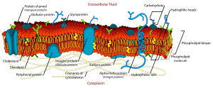
In the cell membrane, a lipid bilayer, including cholesterols (a lipid component) sits between phospholipids to maintain their fluidity at various temperatures and the membrane proteins, including integral proteins, go across the membrane serving as membrane transporters.
Also, the peripheral proteins that loosely attach to the outer (peripheral) side of the cell membrane, acts as enzymes shaping the cell. Since the cell membrane controls the movement of substances in and out of cells and organelles, it is selectively permeable to ions and organic molecules.
Moreso, there are different types of cell membranes in different cell types. The cell membrane, generally, has a lot of cholesterol in it as its lipid component. Although this is different from certain other membranes from within the cell.
However, there are different plants and microbes, like bacteria and algae, which have different protective mechanisms. They have a cell wall outside of them, and the cell wall is tougher and more structurally sound than a plasma membrane or cell membrane.
An animal cell is like a system of membranes that divide it into numerous compartments. Hence, membranes inside the cell surround varieties of organelles. Many cellular functions take place in the membranous organelles. Moreso these internal membranes share many structural features of plasma membranes and are sites for many of the cell’s enzymatic reactions and internal communication systems.
For instance, the nucleus that contains the genetic material of the cell is surrounded by a double membrane with large pores that allow the exchange of materials between the nucleus and cytoplasm.
The outer nuclear membrane is an extension of the membrane of the endoplasmic reticulum, that synthesizes the lipids for all cell membranes. Also, proteins are synthesized by ribosomes that are either attached to the endoplasmic reticulum or suspended freely in the cell contents.
The mitochondria, which are the oxidizing and energy-storing units of the cell, have an outer membrane readily permeable to many substances, and a less-permeable inner membrane studded with transport proteins and energy-producing enzymes.
What does the cell membrane do?
A plasma membrane, however, acts as a selective gatekeeper for the entrance and exit of many substances during cell metabolism. As it transports nutrients into the cell and also transports toxic substances out of the cell, some substances can pass through the membrane with ease and some cannot.
Some substances enter slowly with difficulty whereas some cannot enter at all. This is because conditions outside the cell are different and more variable than conditions within the cell. Hence it is important that the passage of substances across the membrane is rigorously controlled. However, there are 3 main ways that a substance enters across the plasma membrane:
- By a mediated transport system in which the substance binds to a specific site on a transmembrane protein that assists it across the membrane.
- Diffusion along a concentrated gradient
- By endocytosis, in which the substance is enclosed within a vesicle that forms from the membrane surface and detaches inside the cell.
Additionally, cell membranes are involved in a variety of cellular processes such as cell adhesion, ion conductivity, cell signaling and serve as the attachment surface for several extracellular structures, including the cell wall, the carbohydrate layer (glycocalyx), and the intracellular network of protein fibers (cytoskeleton). However, in the field of synthetic biology, cell membranes can be artificially reassembled.
Cell Membrane Diagram


Photo Credit: https://micro.magnet.fsu.edu
Plasma Membrane Structure
The cell membrane is extremely thin measuring about 7.5-10 nm which when magnified can be seen to have three layers (Trilaminar appearance). Additionally, It is a thin semipermeable layer that consists of protein and fats. The Plasma membrane is made up of lipids and glycoproteins.
The fluid-mosaic model is the recent concept accepted describing the cell membrane (plasma membrane) structure. A cell membrane appears as two dark lines by electronic microscopy. Each of the lines is approximately 3nm thick at each side of the light zone.
It creates an image as a result of a phospholipid bilayer (2 layers of phospholipid molecules) which are all oriented with their water-soluble (hydrophilic) ends toward the outside and their fat-soluble portions(hydrophobic) toward the inside of the membrane. An important feature of the phospholipid bilayer is that it is fluidlike.
This feature gives the membrane flexibility and allows the phospholipid molecules to move freely sideways within their own monolayer. However, cholesterol molecules are interspersed in the lipid portion of the bilayer.
Making the membrane even less permeable to water-soluble ions and molecules. Thus decreasing membrane flexibility.
Fluid mosaic model
The fluid mosaic model of S. J. Singer and G. L. Nicolson (1972) replaced the earlier model of Davson and Danielli. So, the fluid mosaic model is the recent concept accepted describing the cell membrane (plasma membrane) structure.
According to the fluid mosaic model, the membrane can be seen as a 2-dimensional liquid, in which lipid and protein molecules diffuse more or less easily. Although the lipid bilayers that form the basis of the membranes do form 2-dimensional liquids by themselves, the plasma membrane also contains a large number of proteins, that provide more structure.
However protein-protein complexes, pickets, and fences formed by the actin-based cytoskeleton, and potentially lipid rafts are examples of such structures.
Composition of the cell membrane
The cell membranes contain a variety of biological molecules, most especially lipids and proteins. However, their composition is not stagnant and changes constantly for fluidity, environmental change, and even fluctuate during different stages of cell development.
For instance, the amount of cholesterol in the human primary neuron cell membrane changes, and this change in composition affects fluidity throughout development stages. The following are the biological molecules that the cell membrane consists of:
-
Lipids
Lipids give cell membranes a fluid character, with a consistency like that of light oil. The cell membrane contains 3 classes of amphipathic lipids, which are phospholipids, sterols, and glycolipids. However, the amount of each of these amphipathic lipids depends on the cell type.
Although Phospholipids are the most abundant in most cases, usually contributing to over 50% of all lipids in cell membranes, the glycolipids account for a small amount of about 2% whereas the sterols make up the rest. However, in RBC studies, about 30% of the plasma membrane is lipid.
The composition of plasma membranes is about half proteins and half lipids by weight for the majority of eukaryotic cells.
The fatty chains in phospholipids and glycolipids usually contain an even number of carbon atoms, usually between 16 and 20. However, some organisms have the ability to regulate the fluidity of their cell membranes by altering lipid composition. This feature is called homeoviscous adaptation.
The phospholipid molecules in the cell membrane are in the liquid crystalline state under physiological conditions. This means the lipid molecules are free to diffuse and exhibit rapid lateral diffusion within the layer they are present in. Actually, the exchange of phospholipid molecules between intracellular and extracellular leaflets of the bilayer is a very slow process.
However, Lipid rafts and caveolae are examples of cholesterol-enriched microdomains in the cell membrane. The fraction of the lipid in direct contact with integral membrane proteins, that are tightly bound to the protein surface is called annular lipid shell and it behaves as a part of the protein complex.
In animal cells, cholesterol is usually dispersed in the irregular spaces between the hydrophobic tails of the membrane lipids, where it gives a stiffening and strengthening effect on the membrane. Also, the amount of cholesterol in biological membranes varies between organisms, cell types, and even individual cells.
Cholesterol is a major component of animal cell membranes that regulates the fluidity of the overall membrane. This means that cholesterol controls the amount of movement of the various cell membrane components based on their concentrations.
Furthermore, in high temperatures, it inhibits the movement of phospholipid fatty acid chains and causes reduced permeability to small molecules and membrane fluidity. However, the reverse is the case in cooler temperatures.
The cholesterol production and concentration are increased in response to cold temperature. At cold temperatures, cholesterol interferes with fatty acid chain interactions, and acting as antifreeze, it maintains the fluidity of the membrane. This is why cholesterol is more in cold weather animals than warm weather animals and sterols perform the same function as cholesterol in plants that lack cholesterol.
-
Carbohydrates
The cell membranes contain carbohydrates too, most especially glycoproteins, but with some glycolipids (cerebrosides and gangliosides). Long carbohydrate molecules attach to proteins at the exterior surface outside of the plasma membrane.
Carbohydrates are very important in the role of cell-cell recognition in eukaryotes. The carbohydrates are located on the surface of the cell where they recognize host cells and share information.
Glycosylation occurs on the extracellular surface of the plasma membrane and glycocalyx is an important feature in all cells, especially epithelial with microvilli. The glycocalyx participates in cell adhesion, and lymphocyte homing. However, galactose is the penultimate sugar and the terminal sugar is sialic acid. Sialic acid carries a negative charge that gives an external barrier to charged particles.
-
Proteins
The cell membrane has a large content of proteins, usually around 50% of membrane volume which are important for the cell. These proteins are responsible for various biological activities. Membrane proteins consist of 3 main types:
- The integral proteins or transmembrane proteins: Examples are Ion channels, proton pumps, and G protein-coupled receptors. Ion channels allow inorganic ions to diffuse down their electrochemical gradient across the lipid bilayer through hydrophilic pores across the membrane. Nerve cells are controlled by ion channels and Proton pumps are protein pumps in the lipid bilayer that allow protons to travel through the membrane by transferring from one amino acid side chain to another. The G-protein coupled receptors are used in cell-to-cell signaling, regulation of the production of cAMP, and the regulation of ion channels.
- Peripheral proteins: Examples are some enzymes and hormones.
- Lipid-anchored proteins: An example is G proteins.
The lipid bilayer of the plasma membrane
A lipid bilayer is a double layer of a phospholipid, cholesterol, and glycolipid molecules that contain chains of fatty acids.
They determine whether a membrane is formed into long flat sheets or round vesicles. The lipid layers are formed through a process of self-assembly. Cell membranes consist mainly of a thin layer of amphipathic phospholipids that spontaneously arrange so that the hydrophobic tail regions are isolated from the surrounding water.
This happens as the hydrophilic head regions interact with the intracellular (cytosolic) and extracellular faces of the resulting bilayer. However, this forms a continuous, spherical lipid bilayer.
The hydrophobic effect (hydrophobic interactions) are the major driving forces in the formation of the lipid bilayers. In fact, an increase in the interaction between hydrophobic molecules allows water molecules to bond more freely with each other, increasing the entropy of the system. This complex interaction can include noncovalent interactions like Van der Waals, electrostatic, and hydrogen bonds.
The phospholipid bilayer structure with specific membrane proteins is responsible for the selective permeability of the membrane with passive and active transport mechanisms. In the lipid bilayer are large proteins that transport ions and water-soluble molecules across the membrane.
Generally, lipid bilayers are impermeable to ions and polar molecules.
The hydrophilic heads and hydrophobic tails arrangement of the lipid bilayer prevent polar solutes from diffusing across the membrane. Although it allows the passive diffusion of hydrophobic molecules. This gives the cell the ability to control the movement of these substances through transmembrane protein complexes such as pores, channels, and gates.
Some proteins in the cell membrane form open pores. These pores are called membrane channels that allow the free diffusion of ions in and out of the cell. Whereas, others bind to specific molecules on one side of a membrane and transport the molecules to the other side.
Membrane structures
The cell membrane can form several types of supramembrane (i.e a membrane placed above another) structures. These structures are responsible for cell adhesion, communication, exocytosis, and endocytosis. Such structures are:
- Caveola: are a special type of lipid raft which are small (50–100 nanometer) invaginations of the plasma membrane in many vertebrate cell types.
- Postsynaptic density: is a protein-dense specialization attached to the postsynaptic membrane.
- Podosome: is a conical, actin-rich structure that is found on the outer surface of the plasma membrane of animal cells. They range from approximately 0.5-2.0 µm in diameter.
- Invadopodium: is an actin-rich protrusion of the plasma membrane, similar to podosomes. However, invadopodia are associated with degradation of the extracellular matrix in cancer invasiveness and metastasis.
- Focal adhesions: These are large macromolecular assemblies or sub-cellular structures that mediate the regulatory effects (signaling events) of a cell in response to extracellular matrix (ECM) adhesion.
- Different types of cell junctions: Cell junctions are a class of cellular structures that consist of multiprotein complexes which provide contact or adhesion between neighboring cells or between a cell and the extracellular matrix in animals.
They are composed of specific proteins like integrins and cadherins. These supra membrane structures can be visible with electron microscopy or fluorescence microscopy.
Membrane polarity
The apical membrane of a polarized cell is the surface of the cell membrane that faces inward to the lumen. This is particularly seen in epithelial and endothelial cells, but also describes other polarized cells like neurons.
The basolateral membrane of a polarized cell is the surface of the plasma membrane that forms its basal and lateral surfaces.
However, the basolateral membrane is a compound phrase referring to the terms basal (base) membrane and lateral (side) membrane which are identical in composition and activity, especially in epithelial cells. The membrane faces outwards, towards the interstitium, away from the lumen.
Moreso, proteins (such as ion channels and pumps) are free to move from the basal to the lateral surface of the cell or vice versa in accordance with the fluid mosaic model.
Epithelial cells are joined by tight junctions near their apical surface to prevent the migration of proteins from the basolateral membrane to the apical membrane.
The cytoskeleton of the cell membrane
The cytoskeleton is found underlying the cell membrane in the cytoplasm. The cytoskeletal elements interact extensively and intimately with the cell membrane.
Cytoskeleton anchors proteins and restricts them to a particular cell surface. For instance, is the apical surface of epithelial cells that line the vertebrate gut and limits how far they may diffuse within the bilayer.
Also, the cytoskeleton has the ability to form appendage-like organelles like cilia, which are microtubule-based extensions covered by the cell membrane, and filopodia, which are actin-based extensions. These appendage organelles are ensheathed in the membrane and project from the surface of the cell in order to sense the external environment and make contact with the substrate or other cells.
The apical surfaces of epithelial cells are dense with microvilli (actin-based finger-like projections) which increase cell surface area. Hence, increasing the absorption rate of nutrients. However, localized decoupling of the cytoskeleton and cell membrane results in the formation of a bleb.
Permeability
The permeability of the membrane is the rate of passive diffusion of molecules through the membrane. However, the permeability of the membrane depends solely on the electric charge, polarity of the molecule, and to a lesser extent the molar mass of the molecule.
Hence, because of the cell membrane’s hydrophobic nature, small electrically neutral molecules pass through the membrane more easily than charged, large ones. Therefore, the inability of charged molecules to pass through the cell membrane results in pH partition of substances throughout the fluid compartments of the body.
Variations of the cell membrane
Most importantly, the cell membrane has different lipid and protein compositions in distinct cell types. Therefore they may have specific names for certain cell types. For instance:
- Sarcolemma in muscle cells: The cell membrane of muscle cells is named Sarcolemma. The average sarcolemma is 10 nm thick compared to the 4 nm thickness of a general cell membrane. Even though it is similar to other cell membranes, sarcolemma has other functions that set it apart, e.g the sarcolemma transmits synaptic signals. This helps generate action potentials and is very involved in muscle contraction. It also makes up small channels called T-tubules that pass through the entirety of muscle cells.
- Oolemma in oocytes: The cell membrane of oocytes is the Oolemma. Oocytes are immature egg cells. Oolemma lacks a bilayer and does not consist of lipids. Instead, the structure has an inner layer (fertilization envelope). Plus the exterior is made up of the vitelline layer, which is made up of glycoproteins. However, channels and proteins are present still for their functions in the membrane.
- Axolemma: The axolemma is the specialized plasma membrane on the axons of nerve cells that is responsible for the generation of the action potential. This membrane consists of a granular, densely packed lipid bilayer that works closely with the cytoskeleton components (spectrin and actin). These cytoskeleton components can bind to and interact with transmembrane proteins in the axolemma.
What is the function of the cell membrane?
The primary functions of the cell membrane can be summarized into the following:
Cell Membrane Functions
- The cell membrane has lipids and glycoproteins that function mainly as receptors and channels that allow specific molecules (ions, nutrients, wastes, and metabolic products) to pass between organelles and also between the cell and the outside environment.
- It helps to keep toxic substances out of the cell and control homeostasis.
- The cell membrane gives the cell a form by anchoring the cytoskeleton to provide shape to the cell.
- It attaches to the extracellular matrix and other cells to hold them together to form tissues.
- Cell membranes surround the cytoplasm of cells and separate the intracellular components from the extracellular environment, enclosing and protecting the cell’s organelles.
- It also separates vital but incompatible metabolic processes carried out within organelles.
- As selectively permeable, the cell membrane is able to regulate what enters and exits the cell. Thus facilitating the transport of materials needed for survival.
Since the cell membrane works as a selectively permeable layer, there are a number of transport mechanism that involves the plasma membranes. Transport mechanism like:
-
Diffusion and Osmosis
Diffusion is the movement of molecules from an area of higher concentration to an area of lower concentration. Thus tending to equalize the concentration throughout the area of diffusion. If a living cell surrounded by a membrane is immersed in a solution having a higher concentration of solute molecules than the fluid inside the cell. Then, a concentration gradient will instantly exist between the two fluids across the membrane.
Cell membranes are selectively permeable. They are permeable to water but variably permeable or impermeable to solutes. However, in free diffusion, it is this selective permeability that regulates molecular traffic.
Hence gases, urea, and lipid-soluble solutes (fats, alcohol, and fatlike substance) are the only solutes that can diffuse through biological membranes with ease and any degree of freedom. Sugars, macromolecules, and many electrolytes move across the membranes through channels or by carrier-mediated processes.
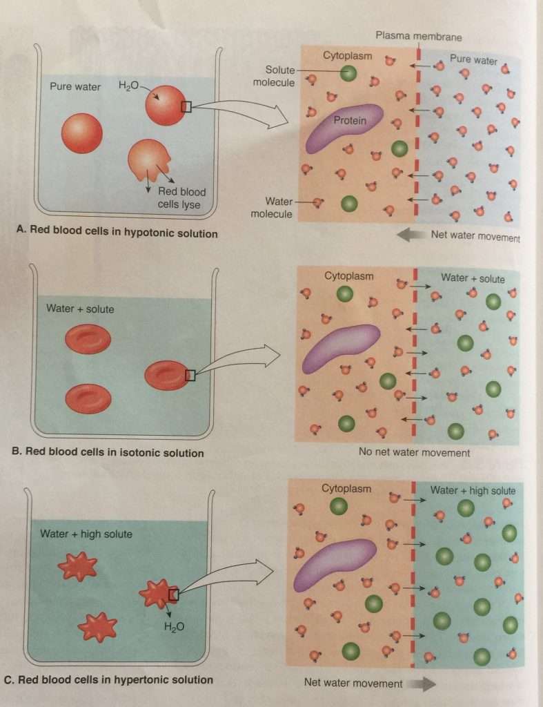
Osmosis is the diffusion of water molecules from an area of high concentration to an area of less concentration. Water flows across the plasma membrane through osmosis because the cytoplasm and external environment are usually at differing concentrations. The process of osmosis can easily be demonstrated with red blood cells.

A: Red blood cells are placed in a beaker of pure water. Water molecules move into the red blood cells through the plasma membrane. Moving from an area of high concentration to an area of lower concentration. The red blood cells swell and lyse.
B: Red blood cells are placed in a beaker of an isotonic solution. There is no net water movement because the concentration of water is equal on each side of the membrane.
C: Red blood cells are placed in a hypertonic solution. The concentration of water molecules is now higher inside the cells. So water moves out of the cells into the beaker and the cells shrink.
Photo credit: Image from Integrated Principles of Zoology (Fifteenth Edition) by Hickman, Roberts, Keen, Eisenhour, Larson, I’Anson. Pg 46
However, water and dissolved ions cannot diffuse through the phospholipid component of the cell membrane since they are charged. So they pass through specialized channels created by transmembrane proteins. Hence, ions and water move through these channels by diffusion. These gated channels may be chemically-gated ion channels, voltage-gated ion channels, or mechanically-gated ion channels.
-
Carrier-mediated transport
Some molecules are of importance to the cell. As it is essential that they enter and leave the cell (e.g sugar and amino acid). However, the plasma membrane is an effective barrier to the free diffusion of most of these molecules of biological significance.
Hence, such molecules are moved across the membrane by transmembrane proteins called transporters or carriers. These transporters enable solute molecules to cross the phospholipid bilayer. They are quite specific by transporting only a limited group of chemical substances or just a single substance at times.
There are 2 distinct kinds of mediated transport mechanism:
- Facilitated diffusion or facilitated transport: The transporter assists the molecule to diffuse through the membrane that it cannot penetrate. Facilitated diffusion supports movement only in the direction of the concentration gradient. It requires no metabolic energy to drive the transportation system. E.g facilitated diffusion aids the transport of glucose (blood sugar) into body cells. The body cells use it as a primary source for the synthesis of ATP.
- Active transport: Energy is supplied to the transporter to transport molecules in the direction opposite to a concentration gradient. Here, molecules are moved against the forces of passive diffusion. E.g is the active transport system that maintains sodium and potassium ion gradients between cells and the surrounding extracellular fluid or external environment.
-
Endocytosis
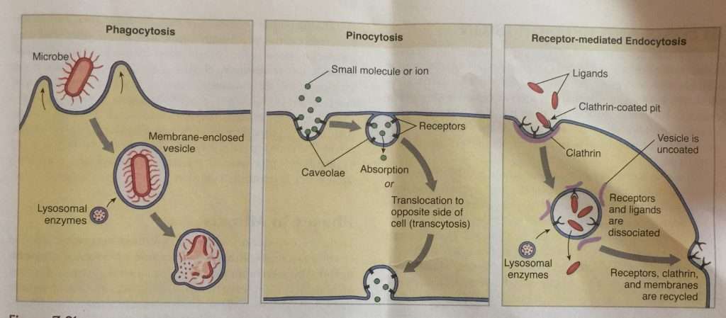
Endocytosis is the ingestion of materials by cells. It is a collective term that describes 3 similar processes: phagocytosis, pinocytosis, and receptor-mediated endocytosis.

In phagocytosis, the cell membrane binds to a large particle and extends to engulf it, forming a membrane-enclosed vesicle, a food vacuole, or phagosome.
In pinocytosis, small areas of the cell membrane, bearing specific receptors for a small molecule or ion, invaginate to form caveolae.
Receptor-mediated endocytosis is a mechanism for selective uptake of large molecules in clathrin-coated pits. The binding of the ligand to the receptor on the surface membrane stimulates the invagination of pits. Lysosomes fuse with the vesicle created during phagocytosis and receptor-mediated endocytosis. lysosomal enzymes digest vesicle contents that are then absorbed into the cytoplasm by diffusion or carrier-mediated transport.
Photo credit: Image from Integrated Principles of Zoology (Fifteenth Edition) by Hickman, Roberts, Keen, Eisenhour, Larson, I’Anson. Pg 49
-
Exocytosis
Since materials can be brought into a cell by membrane invagination and the formation of a vesicle. Exocytosis, however, involves the membrane of a vesicle fusing with the cell membrane and extruding its contents to the surrounding medium.