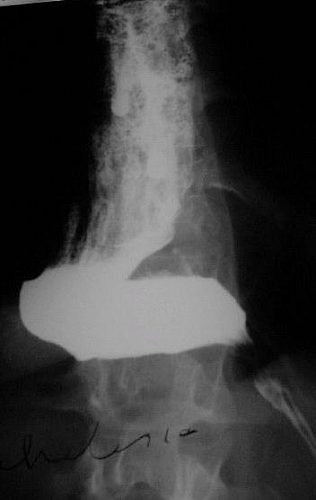
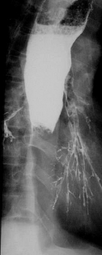
Barium swallow test is a form of imaging in which a patient swallows a liquid medium that can be used for viewing the esophagus and the stomach. A barium swallow study helps to outline the any abnormality along the esophagus or in the stomach.
Table of Contents
Uses/Indications for Barium Swallow in diagnoses of Disease Conditions
Pharyngeal pouch
This is a posterior mucosal protrusion arising in the upper neck just above the cricopharygeus muscle. The patient presents with dysphagia and regurgitation of food on lying down. Plain films may show a fluid level in the pouch but barium swallow is diagnostic with barium filling the pouch which is seen best in the lateral projection when the neck connecting it to the oesophagus can be seen.
Barium swallow for Achalasia
This is a functional disorder of motility resulting in inability of the lower oesophageal sphincter to dilate. The oesophagus becomes widened, sometimes becoming so large that it shows on a plain film of the chest as a mediastinal mass. Food stagnates in the oesophagus; there is regurgitation of food eaten some time previously with dysphagia & weight loss. Barium swallow shows gross oesophageal dilatation with tortuosity, the lower end usually lying horizontally, tapering down to the sphincter, which fails to open. The so-called cigar shape. The oesophagus contains food residue showing as a mottled appearance to the barium.
Barium swallow in diagnosis of Hiatus Hernia
This a protrusion of a portion of the stomach through the oesophageal hiatus of the diaphragm into the chest may range in size from a very small transient hernia to a thoracic stomach. They may be classified as sliding or paraoesophageal. A sliding hernia is the commonest type and occurs when the gastro-oesophageal junction (cardia) and part of the stomach slip upwards above the diaphragm. It is associated with reflux and is usually reducible unless very large. If small, it may only be demonstrated in certain positions. When transient it may not be demonstrated on barium meal at all. With a paraoesophageal hernia, the cardia remains in the normal position and part of the stomach herniates alongside it. Reflux is not a feature and this type of hernia is often irreducible (incarcerated) in which case the hernia remains permanently above the diaphragm and may show as a mass on the chest X-ray containing a fluid level.
Use of Barium swallow in Oesophagitis
Inflammation of the oesophageal mucosa may be secondary to reflux, infections such as monilia, or due to accidental ingestion of a caustic solution. It causes mucosal irregularity with erosions and sometimes ulcers. Strictures may form. Benign strictures usually has smooth, tapering edges in contrast to a malignant stricture which shows an abrupt change in calibre (shouldering).
Barium swallow technique for diagnosis of Carcinoma
This causes a range of appearances depending on the tumour size and degree of malignancy. It is commonest in the distal third presenting as progressive dysphagia with weight loss. On barium swallow, it may present as a mass resulting in a filling defect in the lumen. If infiltrative, it results in narrowing of the lumen initially. Later there is also mucosal destruction and irregularity of the lumen.
Functional disorders
A wide range of functional disorders occur in elderly patients. There may be disordered contractions resulting in a corkscrew appearance. There may be swallowing difficulties due to neuromuscular incoordination as a result of stroke. There is a danger of barium aspiration in these patients during the investigation. This does not usually cause any serious problems unless the patient is much debilitated or the barium of large amount. Physiotherapy is given if a significant amount of barium is aspirated into the smaller bronchi.
Oesophageal varices
These are venous anastomotic collateral veins, usually resulting from portal venous hypertension or portal vein obstruction. They commonly develop as a result of liver cirrhosis and are usually confined to the lower two thirds of the oesophagus. Endoscopy is the investigation of choice but a barium swallow can delineate the large submucosal veins in many cases. If the oesophagus is distended with barium the bulging varices may flatten against the wall and be hidden by the barium. Varices are best shown on films when the barium has passed but coated the oesophageal mucosa in its collapsed state. They show as serpiginous (worm-like) filling defects.
Side effects (Complications) of Barium Swallow
- Injury to the esophagus or stomach
- Inflammation of the Mediastinum (Mediastinitis)
- Peritonitis due to leakage of barium
- Allergies


