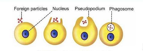
Phagocytosis is the act of ingestion and digestion of microorganisms, insoluble particles, damaged or dead host cells, and cell debris by specific types of cells called phagocytes such as macrophages, neutrophils; Phagocytosis is a form of endocytosis.
The word Phagocytosis is derived from the Greek word-Phagein which means to eat; and it involves the engulfment of large particles like viruses, bacteria, cells, or debris by macrophages and granulocytes in the body.
Table of Contents
Phagocytosis Process
Phagocytosis is the process by which some cells such as Macrophages engulf microorganisms or little solid particles by surrounding the particles or microorganisms using part of their cell membrane; this process is different from Pinocytosis in that Pinocytosis is the ingestion of fluid instead of solid particles.
This part of the cell membrane then separates from the rest of the plasma membrane and forms a free-floating vesicle within the cytoplasm. The membrane bound vesicles, containing solid ingested material are called Phagosomes, and the cells that engulf these particles are called Phagocytes
The process of Phagocytosis was first described and named in 1880 by a Russian zoologist called Elie Metchnikoff who described how the white blood cells ingest foreign particles. He was one of the first scientists to study immunology. Metchnikoff studied the body’s defense against disease-causing agents and invading microorganisms
Phagosomes
Phagocytes are membrane-bound vesicles containing solid materials ingested by Phagocytes (cells that form the vesicles)through a process called Phagocytosis.
Phagocytosis Mechanism
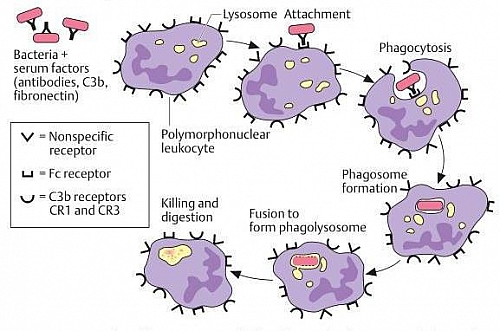
Phagocytosis is initiated when a particle such as a bacterium, a dead cell, or tissue debris binds with receptors on the surface of the phagocyte. Phagocytes then extend their pseudopodia and surround the particles to form phagosomes.
These later fuse with lysosomes to form Phagolysosomes in which the particles are digested. This biochemical mechanism is called respiratory burst. This involves increased oxygen consumption leading to the formation of superoxide ion O2 by NADPH oxidase system.
The superoxide ion subsequently produces hydrogen peroxide (H2O2) which is used for the destruction of the invading microorganism.
Mechanism of Phagocytosis
- When bacteria or foreign body enters the body, first the phagocytic cell sends cytoplasmic extension (pseudopodium) around bacteria or foreign body
- Then, these particles are engulfed and are converted into endosomes like vacuoles. A vacuole is very large and it is usually called the phagosome.
- Phagosome travels into the interior of the cell
- Primary lysosome fuses with this phagosome and forms secondary lysosome
- Hydrolytic enzymes present in the secondary lysosome are activated resulting in the digestion and degradation of the phagosomal contents.
Phagocytosis Steps
Phagocytosis occurs in much the same way as pinocytosis, except that it involves large particles rather than molecules. In the case of bacteria, each bacterium usually is already attached to a specific antibody, and it is the antibody that attaches to the phagocyte receptors, dragging the bacterium along with it. This intermediation of antibodies is called Opsonization.
Steps
- The cell membrane receptors attach to the surface ligands of the particle
- The edges of the membrane around the points of attachment evaginate outward within a fraction of a second to surround the entire particle; then, progressively more and more membrane receptors attach to the particle ligands. All this occurs suddenly in a zipper-like manner to form a closed phagocytic vesicle.
- Actin and other contractile fibrils in the cytoplasm surround the phagocytic vesicle and contract around its outer edge, pushing the vesicle to the interior.
- The contractile proteins then pinch the stem of the vesicle so completely that the vesicle separates from the cell membrane, leaving the vesicle in the cell interior in the same way that pinocytotic vesicles are formed.
Phagocytes Examples
- Macrophages
- Monocytes
- Eosinophils
- Lymphocytes
- Neutrophils
- Dendritic cells in tissues
Phagocytosis Importance and Uses
- Phagocytosis helps to bring in material from outside the cell into the cytoplasm. Pinocytosis is also useful in the same aspect as phagocytosis by helping to ingest fluid into the cytoplasm.
- Phagocytosis helps to clear dead cells from the body
- Protects the body against invading microorganisms thereby involved in the immunity of the body
Inhibition of Phagocytosis
Though phagocytosis was meant to defend the body against invading microorganisms, these invaders sometimes produced chemicals and proteins that also help them to evade being noticed by the immune system and also resist digestion by Phagocytes. Below are the factors that help in achieving Antiphagocytosis
Factors that help microorganisms evade Phagocytosis
- Capsule: some microorganisms are encapsulated and the presence of this capsule renders phagocytosis more difficult. Capsule components may block alternative activation of complement so that C3b is lacking (ligand for C3b receptor of phagocytes) on the surface of encapsulated bacteria. Microorganisms that use this strategy include Streptococcus pneumoniae and Haemophilus influenzae.
- Phagocyte toxins: some bacteria produce toxins that inhibit phagocytic cells from carrying out their functions. Examples of such toxins include Leukocidin from staphylococci and Streptolysin from streptococci.
- Type III secretion system: Macrophages may be disabled by the type III secretion system of certain Gram-negative bacteria, for example, salmonellae, shigellae, yersiniae, and coli bacteria. This type III system is used to inject toxic proteins into the macrophages.
- Inhibition of phagosome: The function of the lysosome can be inhibited by some microorganisms such as tuberculosis bacteria, gonococci, and Chlamydia psittaci.
- Inhibition of the phagocytic oxidative burst: this stops the formation of reactive O2 radicals in phagocytes and renders the phagocytes useless even when they phagocytose the microorganisms. Examples of such microorganisms that inhibit the phagocytic oxidative burst are Legionella pneumophilia and Salmonella typhi.


