Table of Contents
- What is Cellulitis?
- Cellulitis on the face
- Cellulitis on Scalp
- Cellulitis in the Orbit (Orbital Cellulitis)
- Cellulitis of the Neck
- Ludwig’s Angina
- Peritonsillar Abscess (Quinsy)
- Pharyngomaxillary Space Infection
- Tropical Ulcer
- Cellulitis Causes
- How do People get infected with Cellulitis?
- Cellulitis Symptoms and Signs
- Prognosis of Cellulitis
- Laboratory tests (investigations) in cellulitis
- Cellulitis Treatment
What is Cellulitis?
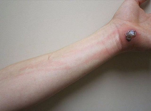
Cellulitis is a non-suppurative, superficial, spreading inflammation of connective tissue. It is a diffuse inflammation of the subcutaneous tissue resulting from invasion by pyogenic bacteria. It spreads along subcutaneous tissues and fascial planes. Streptococcus pyogenes is the commonest cause; less frequently Staphylococcus aureus is responsible and occasionally both species are present creating a symbiosis. The portal of entry may be an invisible puncture, abrasion, insect bite, obvious wounds, fissures, ruptured blister, ulcer or an operative wound. Cellulitis of pelvic origin may be caused by anaerobes and intestinal organisms.
The patient usually complains of pain, redness, swelling and disability. Pain is likely to be severe in sites where the swelling produces tension of anatomically restrained tissues such as the fingers and back of neck. In lax tissue such as the face, pain may be minimal despite gross swelling. The inflamed area throbs with the distension of the pulse and this is aggravated by dependency.
Systemic response to the infection takes the form of toxaemia, malaise and prostration. The degree of systemic disturbance relates more to the nature of the bacterial invader rather than the extent of the lesion. Streptococcal infection causes the greatest reaction. Lymphangitis is evident as tender streaks of discoloured skin.
An area of cellulitis may resolve, leaving apparently normal tissues; it may suppurate with discharge of pus or in severe cases brawny oedema may result in necrosis and tissue replacement by fibrosis. Gangrene may occasionally supervene. Rest, elevation, immobilization of the inflamed part and application of insulating dressings to prevent heat loss are comforting. Antibiotics such as penicillin IV: are useful in the early stages but where suppuration occurs surgical drainage is indicated.
Cellulitis on the face
Cellulitis following a facial wound carries the risk of cavernous sinus thrombosis. Patients should not squeeze or manipulate infected facial foci even if it is small.
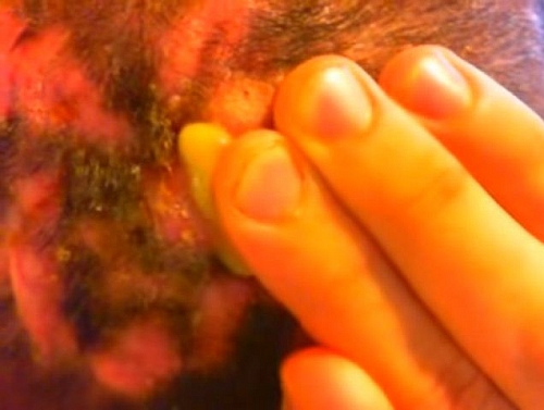
Cellulitis on Scalp
Cellulitis occurs from infection of the subaponeurotic layer of areolar tissue, the pus extending to the attachment of the epicranial aponeurosis, thereby lifting the scalp from the skull. It may lead to necrosis of bone and thrombosis of emissary veins with spread of infection to intra-cranial sinuses.
Cellulitis in the Orbit (Orbital Cellulitis)
This results from wounds around the orbit or spread of infection from the paranasal air sinuses. Proptosis (protrusion of the eyeballs) and paresis (weakness) of ocular movements follow and infection may spread to the meninges or to the cavernous sinus. The eyeball occasionally develops panophthalmitis.
Cellulitis of the Neck
This is usually the sequel to upper respiratory infections, tonsillitis, mastoiditis as well as wounds in the head and neck region. Oedema of the glottis with an ever present danger of asphyxia is a serious complication. Mediastinitis may also occur. Ludwig’s angina and infection of the pharyngomaxillary space are important examples of this form of cellulitis.
Ludwig’s Angina
The characteristic feature of this condition is pronounced intra-oral oedema which accompanies inflammation of the submandibular space. The streptococci responsible for the infection reach the space from the first and second molar teeth. The infection is hemmed in by the attachment of the deep fascia to the hyoid bone and tension rises. The oedematous tongue is displaced upwards and forwards towards the roof of the mouth.
Untreated, the inflammatory exudate spreads along the sheath of the stylohyoid muscle to the submucosa of the laryngopharynx. Sudden death from oedema of the glottis can only be averted by timely surgical intervention.
Cases presenting early may respond to appropriate antibiotic therapy but late cases call for decompression. This is done preferably under local anaesthesia; by means of a curved incision parallel to the lower border of the mandible which divides the deep fascia and the mylohyoid muscles. The wound is then loosely sutured with drainage. To avoid damage to the mandibular branch of the facial nerve, the incision should start at least 2.5 cm below the angle of the mandible.
Peritonsillar Abscess (Quinsy)
This is a complication of acute tonsillitis. The symptoms and signs include: progressive throat pain radiating to the ear, neck rigidity, fever, dysarthria, dysphagia, drooling of saliva, trismus, foul breath and adenopathy. There is also bulging anterior tonsillar pillar and displaced soft palate and uvula from local swelling. The overlying mucosa is inflamed, sometimes with a small spot discharging pus. Differential diagnosis of quinsy includes diphtheria or mononucleosis.
Pharyngomaxillary Space Infection
This closed space is pyramidal in shape with the base upper most at the base of the skull, and the apex at the greater cornu of the hyoid bone. The medial wall is formed by the superior pharyngeal constrictor and the lateral wall successively by the internal pterygoid, the angle of the mandible and the submandibular gland. Streptococcal organisms reach this space from the tonsils and cellulitis here may complicate tonsillectomy or may follow peritonsillar abscess. Painful swallowing, trismus and tender swelling over the lower part of the parotid gland are the symptoms and signs of this infection.
As the space harbours the carotid sheath, delayed decompression is likely to cause jugular thrombophlebitis or erosion of the carotid artery or its main branches. Decompression can be effected through an incision below the angle of the mandible and careful digital exploration of the space.
Tropical Ulcer
This is a form of cellulitis that is produced by Fusobacteria and Borrelia vincenti. There is wide spread tissue necrosis resulting in an extensive ulcer. This may heal satisfactorily under treatment or may require skin grafting; neglected, a chronic indolent ulcer results
Cellulitis Causes
- Common cause by penicillin sensitive Streptococcus pyogenes
- Less frequently caused by Staphylococcus aureus
- May be caused by both streptococcus pyogenes and staphylococcus aureus
- Anaerobic bacteria and gram negative bacteria may cause pelvic infection.
- Fungi and gram negative bacteria may occur in the immunocompromised people with HIV/AIDS, Diabetes Mellitus or Tuberculosis
- Cellulitis associated with the presence of gas in the tissue raises the suspicion of the presence of Clostridium perfringens
How do People get infected with Cellulitis?
- Through punctures
- Abrasion
- Fissures
- Insect bites
- Skin ulcers
- Operative procedures
- Traumatic wound
Cellulitis Symptoms and Signs
- Redness of the affected part of the body such as hand, leg or face
- Pain
- Swelling of the part affected
- Lymphangitis: inflammation of the lymphatic vessels
- Regional Lymphadenitis (swelling of lymph nodes)
- Fever
- Chills
- Feeling of being sick (malaise)
Prognosis of Cellulitis
When left without treatment, cellulitis has different outcomes.
- Resolution may occur
- Suppuration may occur
- The affected tissue dies off (necrosis)
- Fibrosis
- The whole hand or leg may die off (gangrene)
- Septicaemia (infection gets into the blood and many organs will be affected)
Laboratory tests (investigations) in cellulitis
- A full blood count will show multiplication of the white blood cells which points to presence of infection (Leucocytosis)
- Culture of the blood or wound sample will show the specific organism involved
- Blood glucose level may show if the patient has diabetes; other test for tuberculosis and HIV to check if the person is immuno-compromised
Cellulitis Treatment
- Rest and elevation of affected part such as leg or hand
- Analgesics to relief the pains
- Antibiotics to treat the infection
- Surgical drainage if the cellulitis is complicated with pus


